Download
1 / 21
210 likes | 232 Vues
This article discusses the concept of domain exchange in antibodies and its importance in carbohydrate cluster recognition. It explores the structure and function of the 2G12 antibody, which binds to a dense cluster of carbohydrates on the "silent" face of gp120. The study reveals a novel domain-swapped dimer formation in the 2G12 antibody, which allows for high affinity and binding of closely-spaced epitopes. The findings suggest that vaccines mimicking oligomannose clusters may be effective in eliciting protective antibodies.

E N D
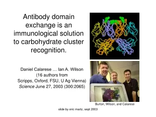
Antibody domain exchange is an immunological solution to carbohydrate cluster recognition. Daniel Calarese … Ian A. Wilson (16 authors from Scripps, Oxford, FSU, U Ag Vienna) Science June 27, 2003 (300:2065) Burton, Wilson, and Calarese slide by eric martz, sept 2003
What is “domain exchange”? Paratope • 2 Ordinary Fabs • VH domains exchanged Forming a Fab dimer with two ordinary VH –VL paratopes + two new VH –VH paratopes Variable Domains Constant Domains H L slide by eric martz, sept 2003
Two Conven- tional Fab’s 2G12 Fab’s “Toober” models slide by eric martz, sept 2003
What is 2G12? • Human monoclonal IgG1 from patient B cells. • Neutralizes unusually broad range of known HIV strains (25-50%). • Binds to dense cluster of carbohydrate epitopes on the “silent” face of gp120. • Unusually high affinity among anti-carbohydrate antibodies. • Antibodies to the 2G12 epitope are unusual in sera from HIV+ patients (by competition). slide by eric martz, sept 2003
The “Silent” face of gp120 • Is an unusual epitope among HIV+ patient antibodies. • Potential protein epitopes beneath the carbohydrate are shielded. • Carbohydrate is made by host (not virus enzymes) hence may be “self” (though the dense cluster of oligomannose residues has not been described on mammalian glycoproteins). • Has multiple glycoforms diluting any one epitope. • Antibodies to carbohydrate epitopes usually have low affinities (Kd micromolar; but 2G12 Kd is nanomolar). slide by eric martz, sept 2003
Why is the structure of 2G12 important? • Paradigm shift: functional framework changes via somatic hypermutation. • Paradigm shift: VH-VH paratope. • Exception to the rule that Fab:antigen interactions are monovalent. • Domain-swapped dimer is unprecedented among >250 published Fab structures. • Explains high affinity and ability to bind closely-spaced epitopes. slide by eric martz, sept 2003
Protein Crystallography • Accounts for 85% of published structures. • Has an overall success rate of ~3%. • <1% of the human proteome has crystallographically known structure. slide by eric martz, sept 2003
Methods • Express full IgG1 in CHO cells. • Papain Fab (completeness confirmed by SDS-PAGE). • Purif. Prot. A/G columns, 30 mg/ml. • 1 μl (ligand 5 to 1 molar ratio) + 1 μl cryst. sol’n (ammonium/sulfate/phosphate, PEG, imidazole malate, pH 6/7), sitting drop crystallization. • Diffract Stanford Synchrotron cryopres. in Liq. N2 • Phase solved by molecular replacement with best-fitting existing Fab structure (1fvd). • Model of known amino acid sequence built into electron density map and refined (with restraints) for best fit. slide by eric martz, sept 2003
Crystallographic Results * ~6,600 atoms slide by eric martz, sept 2003
Framework Somatic MutationsFacilitate Domain Exchange • Weaken VH-VL • Alter H Chain Elbow Conserved ball and socket is not mutated but is dislocated. Mutated residues add stabilization. • Strengthen VH-VH’ See details in Protein Explorer slide by eric martz, sept 2003
Is this bizarre Fab dimer for real?It is not observed in >250 published Fab crystal structures. Fab: about 80% dimer + 20% monomer in solution by • Sedimentation equilibrium analytical ultracentrifugation. • Gel filtration. Intact IgG1 has compact conformation by • Velocity sedimentation coefficient. • Negative staining electron microscopy. • With and without gp140 ligand Apparently the dimer is for real. slide by eric martz, sept 2003
Fig. 2c: Electron microscopy of intact 2G12 IgG1 shows a compact form (neither Y nor T) with or without bound antigen. slide by eric martz, sept 2003
Mutants of 2G12reducing gp120 binding affinity ~100-1,000 fold • Primary combining site (VH-VL) 5/11 • Secondary binding site (VH-VH) 4/5 • Domain exchange-facilitating residues • VH-VH 4/4 • Elbow 3/3 slide by eric martz, sept 2003
Function of HIV oligomannose • DC-SIGN is a human lectin. (Dendritic Cell-Specific ICAM-3 Grabbing Nonintegrin) • Facilitates infection of CD4+ cells by binding HIV carbohydrate. • Speculation: HIV-1 evolved oligomannose to utilize DC-SIGN. • 2G12 exploits this “Achilles heel” of HIV-1. slide by eric martz, sept 2003
Conclusions • 2G12 forms a novel domain-swapped dimer (in 3 crystals and in solution) in both Fab and intact IgG1. • A novel form of paratope occurs at the VH-VH’ interface. • The novel conformation is enabled by somatic mutations to framework residues. • The resulting 4-paratope array accommodates closely spaced carbohydrate epitopes (35 Å, vs. 50-140 Å for Y or T) achieving nanomolar avidity. An epitope on gp120 is predicted. • Vaccines mimicking oligomannose cluster may elicit protective Abs. • 2IG2: scaffold for designing Abs to other epitope clusters? slide by eric martz, sept 2003
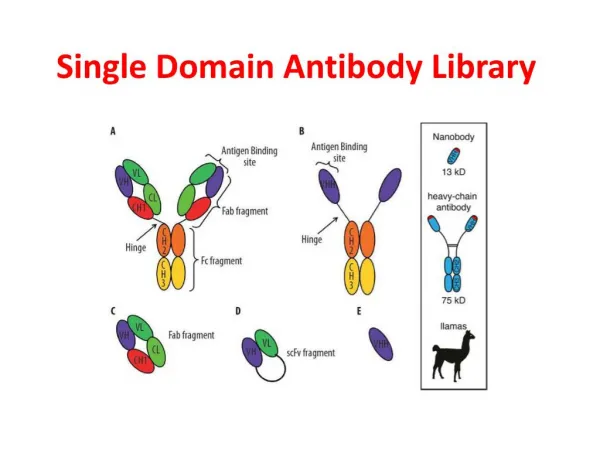
Alternative Antibody Lifestyles Naturally Occurring • Camel H chain dimer (no L; “VHH”) • (Muyldermans et al., TiBS 26:230, 2001) • VH domain-swapped dimer • (Calarese et al., Science 300:2065, 2003) Engineered • Fv (VH-VL with no C domains) • Fv domain-swapped “triabody” (trimer ring) • (Pei et al. PNAS 95:9637, 1997) slide by eric martz, sept 2003
Some High-Impact Crystal Structures • Myoglobin (1960): first example of protein structure. • Lysozyme (1965): first enzyme. • Fab (1973): first immunoglobulin fold (shared in unrelated proteins, first SOD). • tRNA (1975): first RNA structure. • MHC (1987): explained restriction of T cell recognition. • Ribosome (2000): peptidyl transferase is a ribozyme. • 2G12 (2003): unprecedented dimeric paratope with framework somatic mutations. slide by eric martz, sept 2003
First crystal structureswithout big surprises (less impact) • DNA double helix (1973-80): predicted correctly in 1953. • T cell antigen receptor (, 1996). • TCR (2001) • CD1 with phospholipid ligand (2002) slide by eric martz, sept 2003
Fig. 3D slide by eric martz, sept 2003
Fig. 3B slide by eric martz, sept 2003
Fig. 6: Model of 2G12 + gp120 slide by eric martz, sept 2003