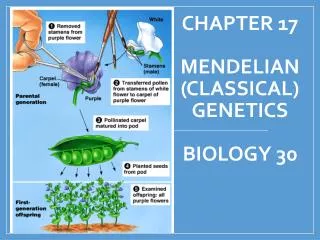
Cell Structure and Function: Exploring the Origin and Processes
300 likes | 408 Vues
Explore the origin of cell theory, key scientists, and understand cell processes from nutrition to reproduction. Learn about the technology that advanced cell studies.

Cell Structure and Function: Exploring the Origin and Processes
E N D
Presentation Transcript
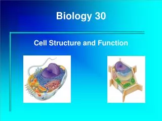
Biology 30 Cell Structure and Function
Unit 1 – Cell Structure and Function In this unit you will learn about the organelles that compose cells, understand how cell transport works, learn about the concepts of diffusion and osmosis as well as their differences, and look at concentration gradientsin cells.
Why study cells? Cells Tissues Organs Bodies bodies are made up of cells cells do all the work of life!
The Origin of Cell Theory Robert Hooke 1665 in England Reported seeing “cells” in thin slices of cork. Coined the term “cell” after the rooms in a monastery. What he saw were not actually cells but the empty walls of dead cells.
The Origin of Cell Theory Antony van Leeuwenhoek Inspired to take up microscopy by having seen a copy of Robert Hooke's illustrated book Micrographia He discovered bacteria, free-living and parasitic microscopic protists, sperm cells, blood cells, microscopic nematodes and rotifers, and much more.
The Origin of Cell Theory Leeuwenhoek is known to have made over 500 microscopes. They were extremely simple devices, using only one lens, mounted in a tiny hole in the brass plate that makes up the body of the instrument.
The Origin of Cell Theory Felix Dujardin 1835 in Germany. Viewed cells under a microscope and discovered that they were filled with a material that is now called protoplasm. Protoplasm is collectively the combination of the nucleus and the cytoplasm
The Origin of Cell Theory Mathias Schleiden 1838 in Germany. He stated that all plants are made of cells. Theodor Schwann 1839 in Germany. He stated that all animals are made of cells.
The Origin of Cell Theory Rudolph Virchow 1859 in Germany. He made three important statements: 1 – Plant cells come only from other plant cells. 2 – Animal cells come only from other animal cells. 3 – Cells come only from other cells.
The Origin of Cell Theory The work of these scientists was used to formulate what modern scientists call “Cell Theory.”
In other words… 1 – All organisms are made up of one or more cells. 2 – Cells are the fundamental functional and structural unit of life. 3 – Cells come only from other cells.
What aided in the development of Cell Theory? The development of microscopes allowed scientists to look more closely at tissues and eventually find similarities between animal and plant ‘cells’ light microscopes were developed 330 years ago and function by passing light through a specimen and bending that light through the use of lenses. resolving power is the clarity of an image (what looks like a star may actually be two stars close together after looking through a telescope) Light microscopes helped discover similarities between plant and animal cells and led to the development of cell theory.
Further Development of Microscopes led to more advanced studies of cells Electron microscopesare able to distinguish objects as small as 2nm in diameter by using beams of electrons. Scanning Electron Microscope (SEM) is used to study the detailed architecture of cell surfaces (uses a metal coating that prevents the beam from going through the cell) Electrons that are emitted create an image of the cell surface.
Transmission electron microscope (TEM) A T.E.M. is used to study internal cell structure. Specimens are cut in thin sections. A beam is aimed through the section and uses electromagnets to magnify and focus the electron beam on a specimen. The picture is then illustrated on a computer screen. Electron microscopes can not look at living specimens because the specimen must be held in a vacuum chamber where all air and liquid are removed.
Cell Processes All life processes involving energy change take place in the cell. This is seen in the many specialized organelles of a cell (for example the golgi apparatus, endoplasmic reticulum, or the nucleolus) that carry out these processes.
Cell Processes Nutrition (eat)– Food molecules supply energy and building materials to the cell. Some cells can make their own food; while other cells take food in from the environment around them. Digestion– Enzymes aid in the digestion and breakdown of complex molecules. Absorption– Cells absorb ions, water, and food molecules from their environment.
Cell Processes Biosynthesis (build molecules)– The process through which cells produce carbohydrates, lipids, and proteins. Respiration (breathe)– Is the exchange of gases between a cell and it’s environment. Cellular Respiration is the release of energy when the bonds of organic molecules are broken. Excretion (remove waste)- Is the process that cells use to expel waste.
Cell Processes Secretion–Cells secrete substances such as vitamins and hormones to cause activities in other cells. Response– Cell activities can be altered in response to outside stimulus such as heat, light, or pressure. Reproduction– Cells divide to form two new cells. In multi cellular organisms (such as you) this results in growth or tissue repair. In single celled organisms (such as an amoeba this is referred to as asexual reproduction.
Cells have 3 main jobs make energy need energy for all activities need to clean up waste produced while making energy make proteins proteins do all the work in a cell, so we need lots of them make more cells for growth to replace damaged or diseased cells The Jobs of Cells Our organellesdo all thesejobs!
A Tour of the Cell Organelle (Latin: small organ) is a cellular structure that’s anatomy gives it a specific role to play in the life of a cell At a minimum, cells must be able to house enough DNA, protein molecules, and internal structures to survive and reproduce Maximum size is limited by the requirement for enough surface area to obtain adequate nutrients from the environment and dispose of wastes
Large cells have more surface area but less surface area relative to the volume of a smaller cell of a similar shape Cells are not all square, rectangular, or circular (muscle and nerve cells can be very long because they are thin and therefore have more surface area per unit volume that spherical cells) Cell size varies with function: nerve cell long to transmit signals quickly, bird eggs nutrients, muscle cells to connect body parts, red blood cell tiny to fit through smallest blood vessels
In cells, macromolecules are suspended in a basic aqueous soup Nucleic acids DNA RNA, mRNA, tRNA Proteins Made up of amino acids. May be conjugated with lipids, sugars or nucleic acids Lipids May be conjugated with other macromolecules. Important constituent of membranes. Carbohydrates Polysaccharides Polysaccharide-protein complexes Glycoproteins Glycolipids
Prokaryotic Cells(Gk: pro-before, karyon-kernal) (e.g. bacteria) lack a nucleus– D.N.A. is coiled into a nucleoid region and thus in direct contact with rest of cell contents. (cytoplasm) Ribosomes in the cytoplasm assemble amino acids into polypeptides, the polymers that make up proteins under direction with D.N.A. The plasma membrane encloses the cytoplasm.
Bacterial cell wall surrounds the plasma membrane and protect the cell, helping to maintain its shape. Some bacteria have a sticky outer layer called a capsule which helps protect and stick to tissues or attach to surfaces. For attachment and movement, bacteria have pili (singular pilus) or flagella (singular flagellum.)
Eukaryotic Cells(Gk: eu-true, karyon-kernal) Partitioned into functional compartments or membranous organelles (nucleus, endoplasmic reticulum, Golgi apparatus, mitochondria, lysosomes, peroxisomes.)
All organelles function together to create and maintain what is called cellular metabolism. Internal membranes provide the surface where many important metabolic processes occur.
Cell size comparison Animal cell Bacterial cell most bacteria • 1-10 microns eukaryotic cells • 10-100 microns • micron = micrometer = 1/1,000,000 meter • diameter of human hair = ~20 microns
Plant Cell Wall Structure Plasma membrane Secondary cell wall Primary cell wall (cellulose) Middle lamella (pectin)