Download
1 / 37
380 likes | 443 Vues
Explore the history and genetic mechanisms behind Agrobacterium tumefaciens in causing crown gall tumors, transferring TIP, and engineering plant cells. Learn about Ti plasmids and virulence functions.

E N D
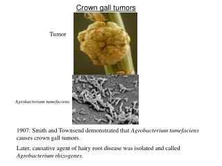
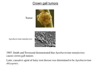
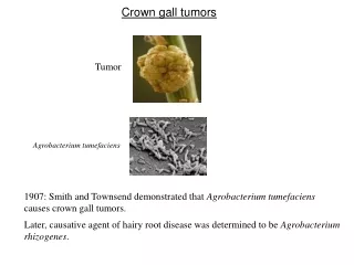
Crown gall tumors Tumor Agrobacterium tumefaciens 1907: Smith and Townsend demonstrated that Agrobacterium tumefaciens causes crown gall tumors. Later, causative agent of hairy root disease was determined to be Agrobacterium rhizogenes.
Agrobacterium genetically engineers plant cells? 1958: Arnim Braun (Rockfeller Univ) showed that tumors could be excised and propagated on in vitro culture media without addition of plant hormones. It was also demonstrated that tumor could propagate even when the bacteria was removed from the tumor. He proposed that the bacteria is able to transfer “Tumor Inducing Principle” (TIP) into plant cells. 1968: Georges Morel (France) found that the tumors released compounds (opines) that Agrobacteria use as nutrients. Therefore, he proposed that (a) bacteria transfer opine synthesis genes into plant cell, and (2) the synthesized opine is transported back into the bacterial cell. This suggests that Agrobacteria are genetic engineer!!! Unacceptable to the scientific community at that time.
TIP is a plasmid 1971 Hamilton and Fall (University of Pennsylvania) reported that a virulent strain of Agrobacterium, when grown at 37oC lost virulence irreversibly. Allen Kerr (Adelaide, Australia) co-inoculated “avirulent” and “virulent” strains on sunflower, and re-isolated the “avirulent” strain. He found that the “avirulent” strain had become virulent. These results indicate that the TIP in Agrobactrium probably resides on a plasmid, which can be transferred between bacterial strains by conjugation.
1974: Ivo Zaenan (University of Ghent) isolated the megaplasmids of Agrobacterium. He called them Ti plasmids, which was later proven to be the TIP. Agrobacterium tumefaciens were classified on the basis of the opine they catabolized such as octopine, nopaline, agropine, mannopine etc. Later it was found that such classification is faulty because most strains catabolize more than one type of opine. Ti plasmids of A6 strain, pTiA6, pTiAch, pTiB6S3 are referred to as octopine Ti plasmids. Ti plasmid of C58 strain (pTiC58) is a nopaline Ti plasmid.
Features of Ti plasmid • About 200 kb. 155 Open Reading Frames (ORFs). • Contains following 5 components: • T region (T-DNA), which codes for sequences that are transferred to plant cell. • The vir region that directs the processing and transfer of T-DNA. • The rep region that is required for the replication of Ti plasmid in bacterial cell. • The tra and trb loci, which direct the conjugal transfer of Ti plasmid between two bacterial cells. • Genes that direct the uptake and catabolism of opines.
vir transfer opine rep Operons in an Octopine Ti plasmid
T-DNA Borders Right: 5’- GXXTGXCAGGATATATXXXXXXGTXAXX-3’ Left: 5’- XGGTGGCAGGATATATXXXXXTGTAAAX-3’ Overdrive is present in Octopine Ti Plasmids
During infection, A. t. carrying an octopine-type Ti plasmid transfers two fragments of DNA to plant cell. These fragments are designated as TL-DNA and TR-DNA and are 13 and 7.8 kb long, respectively. A nopaline type Ti plasmid transfers a single DNA fragment (T-DNA) that is about 20 kb long. TL-DNA and TR-DNA or T-DNA is each flanked by cis-acting 25 bp direct repeats called border sequences (LB and RB or A, B, C and D). The left border is dispensable for T-DNA transfer but right border is essential and acts in polar fashion. Octopine Ti plasmids often contain near RB of TL-DNA another cis-acting element called overdrive, which is required for wild-type transfer efficiency and provides a binding site for a Vir protein called VirC1. Another possible overdrive element is present near RB of TR-DNA but its role is unknown.
LB RB LB RB TL-DNA TR-DNA cleavage • In the presence of Vir proteins, T-region undergoes following processing steps: • Each border is cleaved exactly 4 nt from its left end catalyzed by VirD2 protein, which remains covalently bound to the 5’ end of each cleaved strand. • Bottom strands are recovered as single-stranded (ss) form, referred to as T strand.
Ti plasmid (octopine type) encoded proteins required for T-DNA processing and transfer (vir genes) vir operons: virA, -B, -C, -D, -E, -G. vir F and vir H Vir D1 and D2: D2 is a site-specific endonuclease. Some reports indicate that D1 probably contains topoisomerase I activity, other contradict it. However, both D1 and D2 are required for nicking borders on a supercoiled or relaxed double stranded DNA. Whereas D2 can cleave border sequence on a single stranded DNA without the help of D1. This suggests that D1 could be involved in ripping ds DNA into ss form for D2 to act upon the ss border sequences.
Induction of Virulence Function (initiation of T-DNA transfer) Virulence functions are transcriptionally regulated by 2 component gene regulatory system belonging to a large family of bacterial chemosensors that respond to the chemical environment. Optimal vir gene induction occurs at acidic pH and in the presence of phenolic inducers such as acetosyringone (AS) that are released by wounded plant cells. The vir gene regulatory system operates through two monocistronic virulence genes: vir A and vir G.
Vir A vir A gene is constitutively expressed. Vir A protein is located in the inner membrane and responds to the chemical environment [acidic pH and acetosyringone (AS)]. In the presence of the stimulants, it is auto-phosphorylated. Inner membrane Auto-phosphorylation periplasm cytoplasm linker (pH) kinase AS sensor COOH NH2 receiver Linker responds to pH and interacts with ChvE (a sugar-binding protein encoded by Agrobacterium genome). At sub-optimal AS levels, VirA can be further stimulated by sugars, opines or amino acids.
Vir G vir G gene is also constitutively expressed. Vir G protein is freely available in the cytoplasm. The activated (phosphorylated) Vir A in turn phosphorylates Vir G protein at aspartic acid residue 52. Phosphorylated Vir G becomes the transcriptional activator of the remaining vir genes. Promoters of vir genes possess one of more “vir box” of 12 bp sequence. P P AS P Vir G Vir A A mutant that expresses its vir genes constitutively, contains a vir G mutation called virG-N54D. This mutation leads to a conformation of protein that is similar to phosphorylated Vir G.
T-DNA Processing Vir D1, D2 RB LB Vir D2 RB LB RB LB
Vir C1 and C2 • C1 mutants display lower virulence • C1 binds to the overdrive site. • C2 function is unknown. • It is not clear exactly how binding of C1 on overdrive helps increase the efficiency of T-strand transfer to plants. • Overdrive is absent in nopaline type Ti plasmid.
Vir H or pinF Non-essential. May be involved in detoxification of plant phenolics. VirH exhibits sequence homology with cytochrome P450 like gene. Cytochrome P450 enzymes catalyze NADH-dependent oxidation of aromatic substrates. Vir F Host range factor. Possible interaction with Skp1 proteins to regulate plant cell division cycle. Vir J Putative T-strand binding protein. May have a role in T-strand export from Agrobacterium.
Attachment of Agrobacterium to plant cell • This is a polar two step process: • Mediated by cell associated acetylated, acidic polysaccharides encoded by attR locus. attR mutants are avirulent. This step is reversible because sheer forces are sufficient to dislodge bacteria. • Involves formation of cellulose fibrils by bacterium, which enmeshes large number of bacteria at the wound site. • chvA, chvB and pscA genes are involved in synthesis, processing and export of cyclic beta 1,2 glucans and other sugars, that may be involved indirectly in bacterial attachment. • Plant proteins??? • RAT1 encodes an arabinogalactan protein (AGP). When AGP is blocked by Yariv reagent, transformation is blocked. • RAT4 encodes cellulose synthase like gene
Bacterial attachment Attachment of bacteria to plant cell is a prerequisite for DNA transfer. While many genes involved in attachment process (att genes) have been elucidated, the mechanism of this intriguing process is not fully understood. A 20-kb block of att genes located on bacterial chromosome is involved in attachment. attA1 and attH gene products are probably secreted because attA1 and attH mutation can be complemented by conditioned media (in which plant cell and Agrobacteria were growing). attR gene product is involved in the production of acidic polysaccharides. After initial attachment, Agrobacterium produces a network of cellulose fibrils that bind the bacterium with the plant cell tightly and entraps other Agrobacterium that are not yet attached. Cellulose production is important for efficiency of transformation but not absolutely necessary. Chromosomal virulence genes [chvA (beta-1,2-glucan), chvB (transport protein), pscA] are also involved in the attachment process.
Bacterial attachment Chromosomal virulence genes [chvA, chvB, pscA] are also involved in the attachment process. ChvB is a 235 kDa protein involved in the formation of cyclic β-1,2-glucan. ChvA is a transport protein located in the inner membrane, necessary for the transport of β-1,2-glucan into periplasm. PscA is also involved in the production of β-1,2-glucan.
T-DNA Transfer To The Plant Cell • Until recently the following two topics were among the most debated topics on T-DNA transfer. • Whether T-DNA enters plant cell as ss or ds molecule? Two recent studies categorically demonstrated that T-DNA is ss molecule. • Whether T-DNA travels to the plant cell alone or as a complex with VirE2 protein? Some recent studies have elegantly demonstrated that VirE2 is transported into plant nucleus separately.
T-DNA Transfer Apparatus Encoded by virB operons (11 genes). Each virB except virB1 is essential for tumorogenesis. All 10 VirB proteins have been localized to the inner or outer membrane and most appear either to be integral membrane protein or to be exported from cytoplasm. VirB1 possesses sequence motifs found in transglycosylase and eukaryotic lysozyme, suggesting a role in localized digestion of the peptidoglygan. VirB4 and VirB11 are peripherally bound to others and located primarily in cytoplasm and they contain ATPase activity. Therefore, they may be involved in providing energy for the export of other protein subunits, for T-strand transport or both. VirB proteins constitute a pilus that resembles conjugative pilus and VirB2 is the major subunit of this pilus. VirB7 may help anchor this pilus to the bacterial cell as it is an outer membrane lipoprotein that forms disulfide bonds with the periplasmically localized VirB9.
T-DNA Transfer Apparatus: pilus or pore Cell wall Inner membrane periplasm cytoplasm VirB2 VirB7 VirB4 VirB11 VirB9 VirB1 Other VirB proteins [B3, B5, B6, B8, B10] are minor constituents of the pore. VirB pore
Which Vir Proteins accompany the T-strand into the plant cell? D2 Candidates • VirD2 • VirE2 can bind to ss DNA in vitro • The AcvB protein shares homology with VirJ, which is a ss DNA binding protein. Nopaline strains carrying acvB mutation are avirulent. However, octopine strains are not, probably because this mutation is compensated by virJ gene (which is absent in nopaline strains) • 3. Vir F???
virE genes • Both virE1 and E2 are essential for tumorogenesis i.e. E1 or E2 mutants are avirulent. • VirE2 is ss binding protein and contains nuclear localization signal. • virE2 mutation can be complemented extra-cellularly. • Extracellular complementation is dependent on virB function. • VirE1 is required for transfer of VirE2 but not of T-strand i.e. VirE1 is an export chaperone for E2. • virE2 mutant can be complemented by expressing VirE2 protein • in plant cell, suggesting that VirE2 protein is required in plant cell. VirE3: Transferred from Agrobacteria to plant cell, where it interacts with E2 and facilitates its nuclear import. E2 can interact with VIP1 (a plant protein) for nuclear import. Since VIP1 is a rare protein, Agrobacteria has probably evolved its own counterpart of VIP1, E3, to carry out nuclear transport of E2.
Whether VirE2 travels to the plant cell alone or as a complex with T-DNA? • Argument 1: VirE2 should bind T-DNA in bacterial cell because • It is a strong ss binding molecule and present when T-DNA is generated. • Experimental evidence: induced agrobacteria produce some VirE2 coated T-strands (immunoprecipitation studies). • Argument 2: VirE2 and T-DNA are exported independently into plant cell where E2 binds with T-DNA to prevent nucleolytic attacks. • VirE1, the chaperone of E2 prevents the binding of E2 with T-DNA. • virE2 mutation can be complemented extra-cellularly. • a. by VirE2 protein provided by another strain. • b. by plant cell expressing virE2 gene.
Current model of T-DNA transfer Zupan et al. Plant J. 2000 23(1):11-28
Nuclear targeting of T-DNA in plant cells VirD2 and VirE2 are bound to the T-DNA in plant cell. One or both of these proteins must be responsible for nuclear targeting of T-DNA. VirD2 and VirE2 contain plant-active nuclear localization signal (NLS) sequence. D2 contains two NLSs: one at N-terminal and the other at C-terminal. The latter is probably responsible for targeting. E2 contains two separate NLS regions. ssDNA coated with E2 (when microinjected) can localize into nucleus!! Whereas minus E2 DNA remains in cytoplasm.
Does VirE2 play any role in NLS dependent nuclear targeting? Or does it simply keep T-DNA protected and distended for nuclear import? VirD2 NLS, virE2 mutant can transform plant cell producing VirE2 protein, suggesting that VirE2 NLS is functional.
T-DNA genes carry eukaryotic regulatory sequence 6a 6b 2 1 4 3 5 Nopaline-type T-DNA Octopine-type T-DNA TL 1= tms1 2= tms2 3 = nos or ocs 4= tmr 5= autoregulates synthesis of auxin antagonist. 6b=auxin like?, reduces cytokinin effect? Before the elucidation of T-DNA genes, scientists thought that plant and animal tumors must be biologically similar i.e. the basis of tumorization must be same. For this reason, NIH funded Crown gall studies. But now we know that the two types of tumors are fundamentally different.
Biochemical basis of tumorogenesis: Auxin synthesis Trp IAM IAA tms1= Trp mono-oxygenase tms2= IAM hydrolase tms2 tms1 Cytokinin synthesis Isopentyl adenosine 5’ monophosphate (cytokinin) tmr AMP + Isopentyl Pyrophosphate (IPP) tmr= IPP transferase
Transformation Vectors • ‘Disarmed vectors’: non-tumor inducing Ti Plasmids. • Co-integrative • Binary vectors. Old Agrobacterium binary vectors: low copy New Binary vectors: high copy
Agrobacterium host-range Necrotic response by WHR strains on grape-vines is attributed to VirC activity WHR, LHR, Super-vir strains VirAA6 defect is aggravated in A6 strain suggesting that A6 chromosome produces a repressor that interacts with VirAA6 Agro-infection of maize with MSV Strain infection A6 strain: No C58 Yes C58 + VirAA6 Yes A6+VirAcorrec Yes • Monocots (considered recalcitrant to Agrobacterium infection) • “Agrolistic” system. • Super-binary system: extra set of virG, virC and virB.
“Agrolistic” All major monocots were transformed by biolistic (bombardment of DNA coated gold particles) method by 1992. Agrobacterium produces simple integration patterns, where as biolistic produces complex patterns T-DNA LB RB Co-combardment of binary vector with VirD1 and VirD2 expression cassettes: produced integration patterns just like the ones generated by Agrobacterium.
Transformation Vectors • Basic component: • Selectable markers: nptII, hpt, gentR, bar, EPSPS gene. • Screenable markers: • codon usage, gene shuffling. • Introns • Some fancy stuff: • Scaffold Attachment Region (SAR) or Matrix Attachment Region (MAR): • normalize gene expression, especially of high copy number locus. • Yeast and tobacco SAR were effective, whereas human • and soybean SAR were non-functional. • Site-specific integration system.
Role of the Plant Cell • Transformation efficiency: • Cultivar/ ecotype variation. • Tissue or cell type variation. • Plant factors? Plant genes? • Survey of Arabidopsis T-DNA tagged line. • Root explant transformation. • Germline transformation method.
Efficiency Ecotype/ mutant Block Germ-line transformation Root explant Ws None 87% 0.26 Aa-0 None 89% 0.63 rat-1 bacterial att. 5 0.22 rat-3 bact. Att. 9 0.22 rat5 integration 15 0.22 rat9 integration 6 0.21 rat5 mutant • rat5 mutant contains a T-DNA insertion in histone H2A gene: deficient. • Complemented by overexpression of H2A gene. • Overexpression of H2A improved transformation efficiency • even in WT plants. • rat5 is competent for transient expression but incompetent for • stable expression.
Further Reading: Annu. Rev. Plant Physiol. Plant Mol. Biol. 2000. 51:223–56 AGROBACTERIUM AND PLANT GENES INVOLVED IN T-DNA TRANSFER AND INTEGRATION by Stanton B. Gelvin Plant J. 2000 23(1):11-28 The transfer of DNA from Agrobacterium tumefaciens into plant cell: a feast of fundamental insights by Zupan et al.