Download
1 / 40
460 likes | 954 Vues
Vascular Surgery - 101 Vascular Assessment and PAD. Peter R. Nelson, MD Assistant Professor of Surgery UF College of Medicine Malcom Randall VAMC Deanna Shelpman, RVT Lead Technologist, Vascular Laboratory Shands at UF Medical Center. Objectives. General vascular concepts

E N D
Vascular Surgery - 101Vascular Assessment and PAD Peter R. Nelson, MD Assistant Professor of Surgery UF College of Medicine Malcom Randall VAMC Deanna Shelpman, RVT Lead Technologist, Vascular Laboratory Shands at UF Medical Center
Objectives • General vascular concepts • Vascular history and physical • Vascular diagnostic testing • Demonstration and practical • Shadowing at Shands
Cap (macrophages) Media Core (LDL) Pole (SMC) Lymphocytes AtherosclerosisMature plaque
General Concepts • Atherosclerosis is a systemic disease • Be thorough in both history and PEx • Examine every major arterial system • Synthesize your diagnosis with historical and physical exam clues • Support your diagnosis with studies
History – Risk Factors • Smoking • Diabetes • Hypertension • Hyperlipidemias • Male Gender • Thrombophilias • DVT/PE • Family history
History – Cardiac Disease • Overall 30% of patients presenting with PVD also have severe coronary disease • 31% in aneurysm patients • 26% in patients with cerebrovascular disease • 21% in patients with lower extremity ischemia • Of patients undergoing vascular procedure • 20% have had a previous MI • 7% have had significant CHF • 4% have had previous arrythmia • 60% have abnormal EKG • 7% have had a previous stroke
Lower Extremity PAD Symptoms • Acute Ischemia • 6 P’s • Intermittent Claudication • Thigh/buttock vs. calf • Leriche Syndrome • Classic triad • Critical Limb Ischemia • Rest pain • Tissue loss • Ulceration/cellulitis • Gangrene • Osteomyelitis
Spectrum of Extremity PAD Majority of patients with a decreased ABI or absent pedal pulses are asymptomatic and do not require further evaluation Claudication -perfusion unable to meet demand with exercise Limb Threatening Ischemia -perfusion unable to meet demand at rest
General Physical Exam • Full cardiopulmonary exam • Arrhythmias, murmurs, rubs • Transmitted murmurs may mimic or mask bruits • Full neurologic exam • Cranial nerves • Thorough motor sensory exam • Coordination • MMSE • Ophthalmoscopic exam
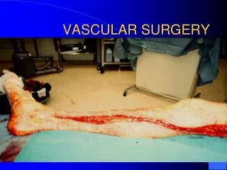
Vascular Exam • Bilateral brachial blood pressures • Palpate pulses • Range 0 - 4+, > 4+ is aneurysmal (note: some people use a 2+ scale) • Carotid, brachial, radial, ulnar, femoral, popliteal, dorsalis pedis, posterior tibial • Ankle-brachial index (ABI) • Listen for bruits • Turbulent flow, more harsh with increasing stenosis, may be lost with critical stenosis • Carotid, subclavian, abdominal (aortic, renal, mesenteric), femoral • Ischemic changes • Shiny skin, hair loss • Dependent rubor, pallor on elevation • Palpate for aneurysms/masses • Aortic, carotid, femoral, popliteal, other
The ABI - Ankle Brachial Index ABI = Ankle systolic pressure (mmHg) Brachial systolic pressure (mmHg) • The ratio of the BEST doppler pressure at the ankle to the BEST brachial pressure • Normal: > 0.96 • Claudication: 0.5 – 0.95 • Rest pain: < 0.3 – 0.5 • Tissue Loss: < 0.3 • Palpable >0.7 • Incompressible >1.3 (without palpable pedal pulses)
Pulse Exam You should be able to draw the arteriogram based on your pulse exam......(and ABIs)
Pulses - Example 1 NORMAL
Pulses - Example 2 R iliac stenosis
Pulses - Example 3 R iliac occlusion
Pulses - Example 4 B iliac disease/L SFA disease (R radial occlusion)
Pulses - Example 5 R SFA occlusion
Pulses - Example 6 R SFA/tibioperoneal disease
Pulses - Example 7 Diabetic - R tiboperoneal disease
Pulses - Example 8 ?? - Medical Student Exam
Pulses - Example 9 Get a new Doppler
Other Diagnostic Studies • Segmental Dopplers/PVRs • 20 mmHg drop across arterial segment • Exercise treadmill testing • 15% drop in ABI • Photoplethysmography • toe pressures • Duplex arterial examination • 2-2.5 X step-up in PSV • Arteriogram • 10 mmHg resting, 20 mmHg induced gradient • CTA/MRA
CW / Continuous Wave Doppler • CW involves a 2 crystal transducer that produces a spectral waveform from the Doppler shift data being received
PVR Waveform • The Pulse Volume Recording (PVR) method measures blood volume changes that occur in the extremities
Photoplethysmography (PPG) Waveform • A PPG waveform is created by the emission of a non-visible infrared light into the skin • Depending on the blood volume in the tissue, more or less light is absorbed and a waveform is created from the backscattered light • Most useful for assessing digital (i.e. toe) pressures/perfusion
Arteriography Aortogram “Runoff”
Demonstration/Practical Questions?