Download
1 / 1
10 likes | 220 Vues
Hemobilia as a Result of Coagulopathy Igor Naryzhny, D.O., Heather Figurelli, D.O., Hymie Kavin, M.D. Department of Medicine, Advocate Lutheran General Hospital Department of Gastroenterology, Advocate Lutheran General Hospital.

E N D
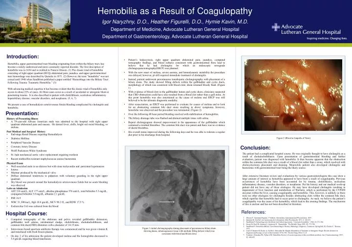
Hemobilia as a Result of Coagulopathy Igor Naryzhny, D.O., Heather Figurelli, D.O., Hymie Kavin, M.D. Department of Medicine, Advocate Lutheran General Hospital Department of Gastroenterology, Advocate Lutheran General Hospital • Patient’s leukocytosis, right upper quadrant abdominal pain, jaundice, computed tomography findings, and blood cultures consistent with gastrointestinal flora lead to believe that he had cholangitis for which an endoscopic retrograde cholangiopancreatography(ERCP) was planned. • With the new onset of melena, severe anemia, and hemodynamic instability the procedure was delayed; however, pt still required immediate treatment of cholangitis. • Instead, patient underwent percutaneous transhepatic cholangiography with placement of a biliary drain. The study showed filling defects within the gallbladder and cystic ducts, morphology of which was consistent with blood clots; drain returned bloody fluid. (Figure 1) • With evidence of blood clots in the gallbladder lumen and cystic ducts, clinicians suspected that CBD obstruction could have also resulted from a blood clot rather than a gall stone. At this point hemobilia was also entertained as the cause of melena and ERCP was still believed to be the ultimate diagnostic modality. • After resuscitation, an ERCP was performed to evaluate for causes of melena and to look for an obstructing common bile duct stone resulting in above symptoms; however, hemobilia was observed and the procedure was terminated. (Figure 2) • Over the following 48 hour period bleeding resolved with stabilization of hemoglobin. • The biliary drainage tube was flushed and drained multiple times with saline. • Repeat cholangiogram showed improvement in the appearance of the gallbladder which contained a residual thrombus. The common bile duct was patent and there was no evidence of ductal thrombus. • His overall status improved during the following days and he was able to tolerate a regular diet prior to his discharge from hospital. Introduction: . Hemobilia, upper gastrointestinal tract bleeding originating from within the biliary tract, has become a widely understood and more commonly reported disorder. The first description of hemobilia was in 1654 and is credited to Francis Glisson. (1) The classic triad of hemobilia consisting of right upper quadrant (RUQ) abdominal pain, jaundice, and upper gastrointestinal tract hemorrhage was described by Quincke in 1871. (2) However, the term “hemobilia” was not coined until 1948 when Sandblom published a paper entitled “Hemorrhage into the Biliary Tract Following Trauma: Traumatic Hemobilia.” (3) With advancing medical expertise it has become evident that the classic triad of hemobilia only occurs in about 22% of cases. (4) Most cases occur as a result of accidental or iatrogenic blunt or penetrating trauma. It is also described in patient with cholelithiasis, acalculous inflammatory hepatobiliary disease, vascular disorders, and neoplasms. (5, 6, 7) We present a case of hemodialysis arteriovenous fistula bleeding complicated by cholangitis and hemobilia. Presentation: • History of Presenting Illness • A 79-year-old African American male was admitted to the hospital with right upper quadrant abdominal pain and nausea. He denied fever, chills, bright red rectal bleeding, or melena. • Past Medical and Surgical History • End-stage Renal Disease requiring Hemodialysis • Diabetes Mellitus • Peripheral Vascular Disease • Coronary Artery Disease • Wolff-Parkinson-White Syndrome • St. Jude mechanical aortic valve replacement requiring warfarin • Recent methicillin-resistant staphylococcus aureus bacteremia • Physical Exam • Well nourished male in no distress but with sinus tachycardia and persistent hypotension • Icterus • Murmur produced by the mechanical valve • Diffuse abdominal tenderness to palpation with voluntary guarding in the right upper quadrant • Dry blood was present around the hemodialysis arteriovenous fistula but no acute bleeding was observed • Labs on Admission • AST 218 unit/L, ALT 177 unit/L, alkaline phosphatase 576 unit/L, total bilirubin 5.3 mg/dL, conjugated bilirubin 5.0 mg/dL, albumin 2.1 gm/dL • INR 16.9 • WBC 31,300/mcL, Hgb 10.0 gm/dL, MCV 90.3 fL, and RDW 17.5 % • Escherichia Coli was cultured from the blood Figure 2 (Blood at Ampulla of Vater) Figure 2 (Blood at Ampulla of Vater) Conclusion: The patient had a complicated hospital course. He was originally thought to have cholangitis as a result of choledocholithiasis. Upon presentation of gastrointestinal bleeding and further evaluation, patient was diagnosed with hemobilia. It then became apparent that the obstruction within the common bile duct was a result of a blood clot rather than a stone, which resolved with cholecystostomy placement and draining. Meanwhile patient also developed cholangitis and bacteremia with gastrointestinal tract being the likely source. After extensive literature review and evaluations by various gastroenterologists this case drew a large amount of interest as hemobilia appeared to have been a result of coagulopathy. Previous occurrences of hemobilia have been accounted for by trauma, cholelithiasis, acalculous inflammatory disease, vascular disorders, or neoplasms. Per his history and our medical work-up patient did not have any of these etiologies. He may have developed cholangitis resulting in impairment of liver function and metabolism of Warfarin, which is performed by the CYP450 enzymes within the liver, causing coagulopathy and hemobilia. This, however, is unlikely as there were no other etiologies for cholangitis besides obstructing clots within the common bile duct, which signifies that hemobilia had to occur prior to cholangitis. As such, we believe the patient’s coagulopathy was the cause of his hemobilia, which lead to the ensuing findings. The mechanism of this is unclear and has not been described in literature. References: Hospital Course: Glisson F: AnatomiaHepatis, 1st Edition. Amsterdam, Janssonium and Weyerstraten, 1654 Quincke H: Ein Fall von AneurysmaderLeberarterie. KlinWochenschr 1871; 8: 349-351 Sandblom P: Hemorrhage into the biliary tract following trauma: ‘Traumatic hemobilia.’ 1948; 24: 571-586 Green M, Duell R, Johnson C, Jamieson N: ‘Haemobilia.’ The British J Surg 2001; 88: 773-786 Sandblom P: Hemobilia (Biliary Tract Hemorrhage): History, Pathology, Diagnosis, Treatment. Springfield, Ill, Charles C. Thomas, 1972 Curet P, Baumer R, Roche A, Grellet J, Mercadier M: Hepatic Hemobilia of Traumatic or Iatrogenic Origin: Recent advances in diagnosis and therapy, review of the literature 1976-1981. World J Surg 1984; 8: 2-8 Yoshida J, Donahue PE, Nyhus LM: Hemobilia: Review of recent experience with a worldwide problem. Am J Gastroenterology 1987; 82: 448-453 • Computed tomography of the abdomen and pelvis revealed gallbladder distension, gallbladder wall edema, intraluminal sludge, cholelithiasis, choledocholithiasis, and common bile duct(CBD) dilatation with a diameter of 14-15 mm. • Intravenous broad spectrum antibiotic therapy was commenced and he was given vitamin K and transfused with fresh frozen plasma. • On day 2 of his admission the patient developed melena and the hemoglobin decreased to 5.4 gm/dL requiring blood transfusion. Figure 1 (initial cholangiography during placement of percutaneous biliary drain showing dense, inhomogeneous tissue with multiple filling defects which was consistent with blood and blood clots)