ABSTRACT
10 likes | 74 Vues
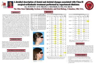
Fig 1: Pt Pre-treatment. Fig 2: Pt Post-treatment with BSSO advancement.

ABSTRACT
E N D
Presentation Transcript
Fig 1: Pt Pre-treatment Fig 2: Pt Post-treatment with BSSO advancement. A detailed description of dental and skeletal changes associated with Class II surgical-orthodontic treatment performed by experienced cliniciansB.L.W.POTTS*, H.W. FIELDS, S. SHANKER, K.VIG, F.M. BECK The Ohio State University, Sections of Orthodontics and Oral Biology, Columbus, OH, USA. RESULTS ABSTRACT The information that details dental changes that accompany pre-surgical and post-surgical orthodontics during orthognathic surgery treatment is disappointing and results in less than ideal surgical change, but is largely derived from university clinic samples and treated by novices. Methods: Seventy-two Class II subjects treated by experienced practitioners (EP) (mean of 26.7 years experience) who underwent surgical-orthodontic treatment with mandibular advancement and rigid fixation were selected and the lateral cephalograms digitized. Mean skeletal and dental values were calculated for pre-treatment, pre-surgical and final treatment time points. Significant effects and differences between means were identified by repeated measures ANCOVA. Treatment efficacy as a percentage of an ideal goal achieved also was calculated. Results: All data showed there were significant positive changes in the position of the mandible. The pretreatment values for the novices and the EP were not equivalent because the novices treated more severe Class II patients. The mean changes demonstrated: For div 1, the upper incisors started proclined and protruded and were retracted and retruded by the novices to near ideal and remained there. The EP did not achieve as much retraction or retrusion. This was a significant difference between the groups. The lower incisors started proclined and protruded in both groups. The novices finished with more proclined and protruded lower incisors. The EP reclined the lower incisors and maintained them as protruded. For div 2, the upper incisors were reclined and retruded. The novices uprighted and protruded them while the EP did a better job of protruding them. The lower incisors were proclined and retruded. Generally, the novice’s became more protrusive and protruded, but not to the ideal. The EP did achieve ideal protrusion. The efficacy data showed no significant differences between the novice and EP groups for any dimensions, but the data were suggestive for div 1 protrusive upper incisor changes. Non-ideal incisor decompensation led to less than ideal final mandibular position. Conclusions: The dental and skeletal changes described by ANCOVA and efficacy analysis showed that pre-surgical orthodontic treatment often does not fully decompensate the incisors, which then limits the surgical outcome even when seasoned practitioners provide treatment. Table 1: Initial (T1): Means, standard deviation for each variable, and ideal values. INTRODUCTION A Table 3: Dental measures at T2 and T3: Means, standard deviaiton, and significant differences between T2-T3 for Div 1 and Division 2 for each variable, and significant differences between Study 1 and Study 2. Non-growing patients with severe Class II skeletal malocclusions benefit from surgical orthodontic treatment to gain functional and facial esthetic improvement. Decompensating the dentition and aligning the teeth to an ideal axial inclination in relation to their basal bone are the goals of pre-surgical orthodontics, but lack of optimal dental decompensation compromises the quality and quantity of the orthognathic surgical correction. The aims of this cephalometric study were to determine if there were differences: 1) In the outcome of incisor inclination between patients with Class II skeletal malocclusions treated by experienced clinicians vs. novice clinicians; 2) At different time points in treatment and 3) Between the measured incisor inclinations and ideal inclinations. DISCUSSION The samples from Study 1 and Study 2 were not comparable at the start of treatment and div 1 and 2 incisor changes were different due to their initial morphology. Generally these differences have been masked in the analysis of means. Because the ANCOVA segregated the div 1 and 2 subjects, a more valid representation of the changes emerged. The results more nearly matched the subsequent efficacy analysis. In both studies the skeletal change was not maximized, resulting in a mild Class II at T3. This can be attributed to the less than ideal pre-surgical decompensation of the dentition. When adjusting for the pretreatment conditions, there were few differences between novices and EP. Preparation and finishing of surgical orthodontic cases requires careful attention to decompensation to avoid the results being a variant of camouflage. D MATERIALS AND METHODS Seventy-two surgical-orthodontic Class II patients were retrospectively selected with the following criteria: consecutively treated Caucasian subjects with Class II molar occlusion, cervical vertebrae maturation of stage 4 or 5 prior to the treatment, and treated with orthognathic surgery of which at least one component was mandibular advancement with rigid fixation. Pre-treatment (T1), pre-surgical (T2), and post-surgical/final (T3) lateral cephalograms were required. Skeletal and dental upper incisor (U1) and lower incisor (L1) measurements for T1, T2, and T3 were recorded and indicated dental compensation and decompensation. Intra-rater reliability was established, and mean and dynamic efficacy evaluations referenced to ideal were calculated. Significant effects and differences between means were identified by repeated measures ANCOVA and efficacy analysis. CONCLUSIONS • Pre-surgically the incisors were not fully decompensated in most patients. • The surgical outcomes were limited by the pre-surgical orthodontic outcomes. • Post-surgically it was necessary to compensate the incisors to gain acceptable occlusion. • The detailed analyses of the efficacy analyses was similar to the ANCOVA analysis because subjects were partitioned by Angle’s occlusal class and division. • The results confirm previous findings of Potts et al.8 with EP practitioners at various geographic locations. Table 2: Skeletal measures at T2 and T3: Means, standard deviation, and significant differences between T2-T3 for Div 1 and Division 2 for each variable, and significant differences between Study 1 and Study 2.


