Download
1 / 81
810 likes | 858 Vues
Explore differential gene expression and genomic equivalence in development, including selective RNA processing and protein modification, through key research findings and mechanisms. Discover the role of transcription factors in tissue-specific gene regulation and chromatin structure modulation. Uncover the significance of epigenetic modifications like DNA methylation in gene expression. Unlock the secrets of genetic regulation for diverse cellular functions.

E N D
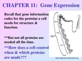
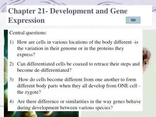
Chapter 2. Differential gene expression in Development Based on the basic assumtion, “Genomic equivalence”, scientist have asked “ how nuclear genes can direct development when these genes are exactly the same in every cell type?” • The answers are • Differentail gene expression • Selective nuclear RNA processing • Selective messenger RNA translation • Differential protein modification
Figure 2.1 Cloning a mammal using nuclei from adult somatic cells Evidence for genetic equivalence -Nuclear transfer and cloning of frog(1952, Briggs and King) -Nuclear transfer from adult frog(1975, Gurdon et al.) -Nuclear transfer in sheep(1997, Wilmut)
Figure 2.2 The kitten “CC” (From 9th Edition) Resurrection is not possible!
Figure 2.4 Nucleotide sequence of the human -globin gene (Part 1)
Figure 2.4 Nucleotide sequence of the human -globin gene (Part 2)
Figure 2.5 Steps in the production of -globin and hemoglobin
Figure 2.6 The bridge between enhancer and promoter can be made by transcription factors
Figure 2.7 The role of the Mediator complex in forming the transcription pre-initiation complex -Mediator complex links the enhancer and promoter to form the initiation complex
Figure 2.7 The role of the Mediator complex in forming the transcription pre-initiation complex (Part 1)
Figure 2.7 The role of the Mediator complex in forming the transcription pre-initiation complex (Part 2)
Figure 2.8 The genetic elements regulating tissue-specific transcription can be identified by fusing reporter genes to suspected enhancer regions of the genes expressed in particular cell types
Figure 2.8 The genetic elements regulating tissue-specific transcription can be identified by fusing reporter genes to suspected enhancer regions of the genes expressed in particular cell types (Part 1)
Figure 2.8 The genetic elements regulating tissue-specific transcription can be identified by fusing reporter genes to suspected enhancer regions of the genes expressed in particular cell types (Part 2)
Figure 2.9 Enhancer region modularity -Enhancer region may have multiple modules for differential gene expression -Each module may need combinatorial association with specific transcription factors for the gene expression
Figure 2.10 Modular transcriptional regulatory regions using Pax6 as an activator
Table 2.1 Some major transcription factor families and subfamilies Pioneer transcription factor: open up the repressed chromatin and maintain activation status
Figure 2.11 Three-dimensional model of the homodimeric transcription factor MITF (one protein shown in red, the other in blue) binding to a promoter element in DNA (white)
Figure 2.12 Pancreatic lineage and transcription factors (Part 1)
Figure 2.12 Pancreatic lineage and transcription factors (Part 2)
Figure 2.14 Chromatin immunoprecipitation-sequencing (ChIPSeq)
Figure 2.14 Chromatin immunoprecipitation-sequencing (ChIPSeq) (Part 1)
Figure 2.14 Chromatin immunoprecipitation-sequencing (ChIPSeq) (Part 2)
Figure 2.15 Chromatin regulation in HCPs and LCPs (Part 1) HCPs are usually found in developmental control genes such as transcription factors HCPs are usually not methylated The default status of HCPs are Open chromatin and the elongation is critical step for gene expression
Figure 2.15 Chromatin regulation in HCPs and LCPs (Part 2) LCPs are usually found in developmental control genes such as transcription factors LCPs are usually methylated The default status of LCPs is inactive form. A specific transcription factor can initiate the gene expression.
Figure 2.21 Model for the regulation of RNA elongation by the Mediator protein Med26
Figure 2.16 Methylation of globin genes in human embryonic blood cells
Figure 2.16 Methylation of globin genes in human embryonic blood cells (Part 1)
Figure 2.16 Methylation of globin genes in human embryonic blood cells (Part 2)
Figure 2.17 DNA methylation can block transcription by preventing transcription factors from binding to the enhancer region
Figure 2.18 Modifying nucleosomes through methylated DNA (Part 1)
Figure 2.18 Modifying nucleosomes through methylated DNA (Part 2)
Figure 2.19 Two DNA methyltransferases are critically important in modifying DNA
Figure 2.20 Regulation of the imprinted Igf2 gene in the mouse
Figure 2.23 Inheritance patterns for Prader-Willi and Angelman syndromes
Figure 15.36 Differential DNA methylation patterns in aging twins
Figure 15.37 Methylation of the estrogen receptor gene occurs as a function of normal aging
Figure 17.19 Cancer can arise (A) if tumor-suppressor genes are inappropriately turned off by DNA methylation or (B) if oncogenes are inappropriately demethylated