
Usefulness of 2-D Ionization Chamber Array for Dynamic IMRT Dose Verification
This study evaluates the dosimetric characteristics of the MatriXX ionization chamber array for dose verification in dynamic IMRT fields with a micro-MLC. Results show good agreement with ionization chamber measurements for 6 MV photon beam parameters and limitations in spatial resolution for certain field sizes. The MatriXX system offers accurate dose measurements but exhibits challenges in steep slope regions due to ionization chamber arrangement. Conclusion emphasizes its utility with caution for high gradient fields.

Usefulness of 2-D Ionization Chamber Array for Dynamic IMRT Dose Verification
E N D
Presentation Transcript
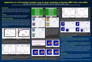
Application of a 2-D ionization chamber array for dose verification of dynamic IMRT with a micro-MLCFujio ARAKI, PhD1, S. TAJIRI2, H. TOMINAGA2, K. KAWASAKI2, K. KOGO2, M. YOSHITANI3, N. ARAKI3, L. Müller41Kumamoto University School of Health Sciences, Kumamoto, Japan, 2Kumamoto Radiosurgery Clinic, 3Toyo Medic Co., Ltd., Tokyo, Japan, 4Scanditronix Wellhöfer GmbH, Schwarzenbruck, Germany (a) (d) (a) Original dose profiles. (b) After transferring to the same grid size. Fig. 5 Comparison of IMRT dose profiles between MatriXX and RTPS for before and after grid transform. The dose profiles of RTPS are transferred to the same grid size as MatriXX. (b) (e) Fig. 3 Comparison of dose profiles between MatriXX and RTPS tested using a bar pattern of mMLC with 3, 4.5, 5.5 and 10 mm wide at isocenter.Measurements were performed at a depth of 10 cm in a solid water phantom for a 6 MV x-ray beam, SSD=95 cm. The dose profile of RTPS is transferred to the same grid size (7.62 mm) as MariXX in figure of lower left. (a) Original dose profiles of MatriXX and RTPS. (b) After transferring to the same grid size. BACKGROUND A pixel-segmented ionization chamber, MatriXX, (Scanditronix Wellhöfer, Germany), has been designed to ease the two-dimensional (2D) verifications of fields with complex shapes like intensity modulated radiation therapy, IMRT. The detector features a 24×24 cm2 active area divided in 1020 independent plane parallel ionization chambers. The sensitive volume of each signal ionization chamber is 0.07 cm3 (4.5 mm diameter×5 mm height) and chamber-to-chamber distance is 7.62 mm). PURPOSE This study aims to examine dosimetric characteristics of MatriXX and its usefulness for dose verification of dynamic IMRT fields with a dedicated Novalis shaped beam radiosurgery unit equipped with a micro-multileaf collimator (mMLC: BrainLAB, Heimstetten, Germany). MARERIALS AND METHOD The absorbed dose for MatriXX was calibrated with a Farmer ionization chamber in a solid water phantom. The MatriXX response was tested as to dose linearity and dose rate dependence. Basic parameters of radiation beam (TPRs, OARs, output factors) were compared with ionization chamber measurements for a 6 MV photon beam. The spatial resolution of MatriXX was tested using a bar pattern of mMLC with 3, 4.5, 5.5 and 10 mm wide at isocenter. The analysis of measured (MatriXX and ionization chamber) and calculated absorbed dose distributions were performed for complex dynamic IMRT fields. The dynamic IMRT dose distribution was calculated with the BrainSCAN 3-D treatment planning system (BrainLAB, Heimstetten, Germany) for stereotactic radiotherapy (SRT) fields formed by mMLC. Fig. 6 Comparison of IMRT dose profiles between MatriXX and RTPS. The dose profiles are evaluated by DD=3% and Dd=3mm of “Gamma method”. RESULTS AND DISCUSSIONThe dose response of MatriXX is linear in the range 0.1-10 Gy. The dose rate dependence is within 0.1% in the range 1.6-8 Gy/min. The TPR and OAR data measured in a 6 MV photon beam is in good agreement with ionization chamber measurements (Figs. 1(a) and 2). Output factors measured with MatriXX and an ionization chamber agree within 1% for field sizes in the range from 1.2×1.2 cm2 to 10×10 cm2 (Fig. 1(b)).The spatial resolution of MatriXX is 7.62 mm, which cannot recognize the bar pattern of 3 mm, as it contains spatial frequencies much higher than MatriXX’s intrinsic resolution (Figs. 3(d) and (e)). The bar pattern of 4.5 and 5.5 mm can be recognized by MatriXX but the dose profile does not contain the full modulation due to limited spatial resolution (Figs. 3(d) and (e)). The MatriXX profile agrees with that of RTPS for the 10 mm bar pattern (Figs. 3(a)~(c)). A direct comparison can be made between dose profiles measured with MatriXX and the calculated dose profiles of RTPS when they are integrated to the same spatial resolution of 7.62 mm (Fig. 4). With this method, excellent agreement can be achieved between planned data and measurement with MatriXX. In dose verification of IMRT fields, MatriXX agrees with those of RTPS in the region where the dose slope is gentle, while it shows some difference from the dose distributions by RTPS in the steep slope regions (Figs. 5 and 6). This is because the dose distribution cannot be faithfully reflected in the linear interpolation in relation to the resolution due to the arrangement of MatriXX ionization chambers (at 7.62 mm intervals). CONCLUSIONSThe pixel-ionization-chamber 2D-MatriXX is found to be able to measure the given dose (absolute dose) and dose distributions simultaneously. It is useful for dose verification of dynamic IMRT pre-treatment. As a word of caution, the limited number of detectors and the dimension of a single detector have to be always kept in mind when dealing with very high gradient or very small field sizes formed with mMLC, but within these limitations it can be still used for the verification of these fields.Reference1. S. Amerio, A. Boriano, F. Bourhaleb, et al., “Dosimetric characterization of a large area pixel-segmented ionization chamber,” Med, phys. 30, 414-20 (2004).2. M. Stasi, S. Giordan, R. Cirio, “D-IMRT verification with a 2D pixel ionization chamber: dosimetric and clinical results in hed and neck caner.” Phys. Med. Biol. 50, 4681-94 (2005). (c) (a) TPR (b) Output factor Fig. 1 TPR and output factor data obtained for a 6 MV x-ray beam using an ionization chamber and MatriXX in a solid water phantom. The output factors for small fields less than 2 x 2 cm2 were measured with a diode detector (SFD). (a) Procedure to compare MatriXX with RTPS. (b) (a) depth=1.5 cm (b) d=20 cm Fig. 2 Comparison of beam profiles measured with an ionization chamber in a water phantom and with MatriXX in a solid water phantom. The irradiations were performed at dmax and 20 cm, and for 4.2 x 4.2 cm2 and 10 x 10 cm2 field sizes in a 6 MV x-ray beam. The results are normalized to the central beam axis. (d) (c) Fig. 4 Procedure to compare fluence map of MatriXX with RTPS’s for IMRT dose verification. Fig. (d) is final dose profiles of MatriXX and RTPS transferred to the same grid size.


