Download
1 / 3
30 likes | 44 Vues
Lymphoma involvement in the lung can occur from hematogenous spread or contiguous invasion from nodal disease, but also rarely forms as primary pulmonary lymphoma (PL). PL has been classified as the World Health Organization into primary pulmonary Non- Hodgkinu2019s lymphoma (PPNHL), primary pulmonary diffuse large B-cell lymphoma and lymphomatoid granulomatosis. Here we report on 2 patients diagnosed with a subtype of PPNHL

E N D
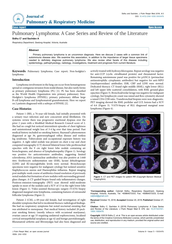
Sidhu and Sachdev, J Pulm Respir Med 2018, 8:5 DOI: 10.4172/2161-105X.1000476 JournalofPulmonary&RespiratoryMedicine ISSN: 2161-105X Journal of Pulmonary & Respiratory Medicine Case report Pulmonary Lymphoma: A Case Series and Review of the Literature Sidhu C* and Sachdev A Open Access Respiratory Department, Geelong Hospital, Victoria, Australia Abstract Primary pulmonary lymphoma is an uncommon diagnosis. Here we discuss 2 cases with a common link of autoimmune disease also. We examine this association, in addition to the importance of larger tissue specimens needed to definitely diagnose pulmonary lymphoma. We also review other facets of this disease including epidemiology, pathophysiology, radiology, investigations, treatment and prognosis from current literature. Keywords: Pulmonary lymphoma; Case report; Non-hodgkin’s lymphoma Introduction Lymphoma involvement in the lung can occur from hematogenous spread or contiguous invasion from nodal disease, but also rarely forms as primary pulmonary lymphoma (PL) [1]. PL has been classified as the World Health Organization into primary pulmonary Non- Hodgkin’s lymphoma (PPNHL), primary pulmonary diffuse large B-cell lymphoma and lymphomatoid granulomatosis. Here we report on 2 patients diagnosed with a subtype of PPNHL [2]. Case Histories Patient 1 (RR), a 76-year-old female, had initially presented with a urinary tract infection and new concurrent atrial fibrillation. On systemic review there was progressive exertional dyspnea over the prior 2 years with a Modified Medical Research Council score of 2. She had no cough but noticed intermittent episodes of chest tightness and unintentional weight loss of 3-4 kg over that time period. Past medical history included no smoking history, Raynaud’s phenomenon diagnosed at age 36, gastroesophageal reflux disease and restless leg syndrome. Tuberculosis and occupational exposure history was negative. A right sided nodule was present on chest x-ray and chest computed tomography (CT) showed bilateral lower lobe peribronchial opacities with the 9 cm right lower lobe nodule containing air bronchograms, and absence of lymphadenopathy (Figure 1). Serology was positive for anticentromere antibodies, suggesting limited scleroderma; ANA (antinuclear antibodies) was also positive at 1:640 titre. Erythrocyte sedimentation rate (ESR), lactate dehydrogenase (LDH) and B2-microglobulin levels were normal. Bronchoscopy specimens were negative for malignant cytology but isolated sensitive Staphylococcus aureus and Haemophilus influenza. Repeat imaging post multiple-week course of antibiotics found resolution of previously noted nodules but formation of new nodules with surrounding ground- glass changes. A CT-guided biopsy found only inflammatory changes. Positron-emission-tomography (PET) scan showed mild-moderate uptake in most of the nodules and a SUV of 13 in the right lower lobe lesion (Figure 1). Video-assisted thorascopic surgery (VATS) biopsy diagnosed marginal zone lymphoma, containing features of extranodal follicular lymphoma (Figure 2). Patient 2 (CM), a 69-year-old female, had investigation of right shoulder symptoms that led to incidental thoracic radiological findings. She had no respiratory symptoms but noted nocturnal sweating that was attributed to menopause. Past medical history included 20 pack- year smoking history (mostly tobacco with occasional marijuana), ovarian cancer at age 33 requiring unilateral oophorectomy, localized cervical intraepithelial neoplasia at age 42 and benign parotidomegaly. Rheumatoid arthritis and fibromyalgia had also been diagnosed and actively treated with hydroxychloroquine. Repeat serology was negative for anti-CCP (cyclic citrullinated protein) and rheumatoid factor. Remaining autoimmune panel was positive for pANCA (perinuclear antineutrophilic cytoplasmic antibodies) but negative for anti-MPO (myeloperoxidase) antibodies. ESR, CRP and LDH were normal. Dedicated thoracic CT found right middle (RML), right lower (RLL) and left upper lobe scattered consolidation, with RML ground-glass opacities (Figure 3). Bronchoalveolar lavage was negative for malignant cytology, but lymphocyte count was raised and flow cytometry showed a raised CD4-CD8 ratio. Transbronchial biopsies were non-diagnostic. PET imaging showed the RML perihilar and LUL lesions had a SUV of 6.6 (Figure 3). VATS-biopsy of RLL diagnosed marginal zone lymphoma (Figure 4). Figure 1: CT and PET images for patient RR (Copyright Barwon Medical Imaging/bmi). *Corresponding author: Calvinjit Sidhu, Respiratory Department, Geelong Hospital, Victoria, Australia, Tel: +60864573333; Fax: +60864573333; E-mail: caljitsid@gmail.com Received October 15, 2018; Accepted October 20, 2018; Published October 27, 2018 Citation:Sidhu C, Sachdev A (2018) Pulmonary Lymphoma: A Case Series and Review of the Literature. J Pulm Respir Med 8: 476. doi: 10.4172/2161- 105X.1000476 Copyright: ©2018 Sidhu C, et al. This is an open-access article distributed under the terms of the Creative Commons Attribution License, which permits unrestricted use, distribution, and reproduction in any medium, provided the original author and source are credited. J Pulm Respir Med, an open access journal ISSN: 2161-105X Volume 8 • Issue 5 • 1000476
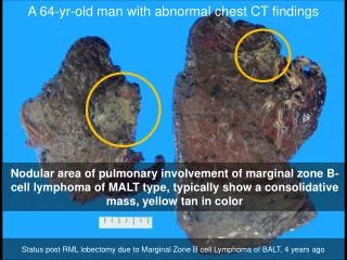
Citation: Sidhu C, Sachdev A (2018) Pulmonary Lymphoma: A Case Series and Review of the Literature. J Pulm Respir Med 8: 476. doi: 10.4172/2161- 105X.1000476 Page 2 of 3 of diagnosis [1,3,4]. These were first described by Bienenstock in 1973, and is estimated as 0.4% of all malignant lymphomas and <1% of primary pulmonary malignancies [2,5,6]. Our cases of PL are a subtype of mucosa-associated lymphoid tissue (MALT) lymphomas. MALT is not present in normal bronchial mucosa but is in the gastrointestinal tract (GIT) [7]. Hence MALT lymphoma is most frequent in the stomach with strong links to Helicobacter pylori infection [8]. They represent 69-78% of extranodal lymphomas [8]. Similar to our 2 cases, most either have non-specific symptoms (cough, fatigue, dyspnea, hemoptysis, diaphoresis, chest pains) or are found as incidental radiological abnormalities [5-7]. <20% were found to have B-symptoms [9]. Crackles during chest auscultation is the most common examination finding [1]. Incidence peaks during the sixth and seventh, just as in our 2 patients, but has no association with female gender [1,3]. Chronic inflammation is thought to be the underlying pathophysiology, from causes such as smoking, infection and connective tissue disease (CTD) [5]. 2/3 of cases were reported to have a significant smoking history. Pulmonary MALT lymphoma is strongly linked with primary Sjogren’s syndrome with a relative risk (RR) of 4-16 [10]. Risk was related to higher rheumatoid factor, lymphopenia, cryoglobulinemia and moderate-to-severe disease activity [10]. Rheumatoid arthritis has a lymphoma RR of 1.5-4 and increased lung cancer risk, which were also associated with CTD severity [11]. Most work has been done in diffuse scleroderma, which found an incidence ratio of 1.41 for developing cancers, but Chatterjee found their limited scleroderma cohort to have much greater cancer risk [11,12]. Lung cancer was the common cancer found. No increased cancer risk has been found with the presence of CTD-interstitial lung disease [13]. Long term immunosuppressant use for CTD are too a recognized risk factor for malignant disease [11]. The inflammation leads to genetic change, with t (11;18) being the most common of 3 translocations [6]. This increase nuclear-factor kappa-beta release to drive antigen-independent B-cell proliferation, which have undergone heavy and light chain rearrangement [6]. They express CD20, CD21, CD35, CD79a and IgM but lack CD5, CD10, CD23 and IgD [1,6]. An important consideration also is rheumatological disease can manifest as a paraneoplastic syndrome, via a similar precipitant causing both malignancy and a separate paraneoplastic process, or paraneoplasia due to cancer toxins or hypersensitivity to antigens post-apoptosis [14]. Atypical features present include rapid symptom onset, unusual age of CTD diagnosis, poor response to CTD treatment, abnormal clinical features (such as joints involved) or abnormal pathology (such as persisting cytopenias/hypergammaglobulinemias) [14]. Kim et al. surmised that “any radiological abnormality in the lung can be lymphoma”, however the findings in our cases: air bronchograms, peribronchial thickening and alveolar opacities, have been seen previously [5,6]. Also has ‘ground-glass with halo’ sign, ‘positive angiogram’ sign and distended bronchi, which is thought to be most specific [4,6]. Sole ground-glass opacities and interlobular septal thickening are atypical [4]. 10% also have pleural effusions, with 34% having hilar lymphadenopathy and 5%-30% having mediastinal lymphadenopathy [4,6,15]. Invasive pulmonary aspergillosis (IPA) is a radiological differential, but Kawel’s comparisons found pleural consolidation had a greater association for this, but lobar consolidation containing air bronchograms being more specific for PL. Pulmonary nodules occurred more in IPA, but were non-specific for either, and tended to resemble the ‘halo’ sign [16,17]. Nodules with spiculated Figure 2: These micrographs show a nodular lymphocytic lesion composed of a mixture of large lymphocytes, centrocyte-like small lymphocytes, monocytoid lymphocytes and small lymphocytes, which expand alveolar septae and fill alveolar spaces (100X and 400X magnifications). There is some sclerosis in the background and residual germinal centres are identified. Courtesy of Dr WW Yong, Department of Anatomical Pathology, Australian Clinical Labs/St John of God Pathology (Geelong). Figure 3: CT and PET images for patient CM (Copyright Barwon Medical Imaging/bmi). Figure 4: The lung architecture is effaced by a population of small lymphocytes forming nodules, some of which have reactive germinal centres (100X and 400X magnifications). Some of these small lymphocytes show clear cytoplasm (monocytoid lymphocytes). The lymphocytic infiltrate expand alveolar septae and fill alveolar spaces. Courtesy of Dr WW Yong, Department of Anatomical Pathology, Australian Clinical Labs/St John of God Pathology (Geelong). Discussion Primary pulmonary lymphoma is defined as lymphomatous proliferations in the lung, lobe or primary bronchus, with or without mediastinal involvement, and no extrathoracic disease within 3 months J Pulm Respir Med, an open access journal ISSN: 2161-105X Volume 8 • Issue 5 • 1000476
Citation: Sidhu C, Sachdev A (2018) Pulmonary Lymphoma: A Case Series and Review of the Literature. J Pulm Respir Med 8: 476. doi: 10.4172/2161- 105X.1000476 Page 3 of 3 margins, or containing cavitation (‘air crescent’ sign) were also more IPA-related [16]. Bronchoalveolar lavage with lymphocytic alveolitis has a 66% association with PL, and greater specificity when B-lymphocyte counts >10% [1]. Cytology can reveal medium lymphoid cells with lymphoplasmacytoid differentiation and irregular nuclei [2]. B-cell clonality is also useful with 95% sensitivity and 97% specificity [6]. Newer polymerase chain replication (PCR) techniques have 83% sensitivity and 94% specificity in detecting associated IgH-gene arrangements, but their current role is in determining recurrence or disseminated disease [1,17]. Fluorescent in situ hybridization (FISH) techniques to detect associated MALT-1 gene rearrangements are being developed [4]. Smaller biopsies should be interpreted with caution, similar to our 1st case, especially if there are atypical features, persistence or progressive lesion change, or sampling technique quality. The development of transbronchial techniques and cryobiopsies could be sufficient in the future by combining morphological examination, newer B-cell clonality testing and cytogenetic analysis, but currently larger specimens via VATS or thoracoscopy are preferred. Differential diagnoses to consider include infectious pneumonia, tuberculosis, lepidic pneumonia, eosinophilic pneumonia, sarcoidosis, cryptogenic organizing pneumonia, hypersensitivity pneumonitis, alveolar hemorrhage, vasculitis, pseudotumor, Castleman’s disease, lung plasmacytoma or alternative malignant disease [2,3]. Post-diagnosis investigations recommended include completion staging CT and bone marrow biopsy, to exclude marrow involvement present in 20%-30% of MALT lymphomas [2,3]. Ear-nose-throat, ophthalmologic and upper GIT evaluation should be considered [1- 3]. PET scans previously had an indeterminate role in PL but Beale found 80% were significantly avid despite their low-grade nature, as can be seen with our testing [6,18]. This is further improved with combined CT-PET modalities. They also have greater relevance in non-gastric MALT lymphoma by confirming higher stage disease at diagnosis or identifying disease progression and relapse during follow- up [19]. Observation has been suggested for asymptomatic PL given likely indolence [5]. Surgical resection can aid both in diagnosis and as potential curative treatment but has yet to show significant survival advantage [7]. Chemotherapy is favoured to avoid (post) surgical complications, or when lung function is impaired or to avoid post- resection recurrence [20]. Various chemotherapy trials have not identified a superior regiment, but the use of rituximab could become more standard. Histology is the main prognostic factor for PL, but B-symptoms can also signal worse disease course [4,6,20]. Overall survival (OS) has been linked with Ann-Arbor stage, number of extranodal sites involved and intermediate-to-high International Prognostic Index scores [4]. 15% of MALT lymphomas transform to alternative significant hematological disease [6]. Survival at 3,5 and 10 years have been quoted as 86%, >80% and 50%-70% respectively [3,5,7]. Risk of relapse is 50% at 2 years and 35% at 5 years [1]. A current prognostic marker in development is the Tumor Microvascular Density, a measure of lymphoma neovascularization [4]. Conclusion In summary, PL is rare but does have concerning malignant potential given risk of transformation and recurrence. Radiology and bronchoscopy findings are non-specific, raising the importance of sufficient tissue samples for definitive histological diagnosis. Cancer occurrence in CTD is an important consideration. Acknowledgements 1. We thank Dr Wei Wei Yong and Department of Anatomical Pathology, Australian Clinical Labs/St. John of God Pathology (Geelong) for assistance with histopathology images and description. 2. We thank Barwon Health and Eastern Health (Victoria) library staff for assistance with literature searches and obtaining references. References 1. Cadranel J, Wislez M, Antoine M (2002) Primary pulmonary lymphoma. Eur Respir J 20: 756-762. 2. Poletti V, Ravaglia C, Tomassetti S, Gurioli C, Casoni G, et al. (2013) Lymphoproliferative lung disorders: Clinicopathological aspects. Eur Respir Rev 22: 427-436. 3. Graham B, Mathisen DJ, Mark EJ, Takvorian RW (2005) Primary pulmonary lymphoma. Ann Thor Surg 80: 1248-1253. 4. Cardenas-Garcia J, Talwar A, Shah R, Fein A (2015) Update in primary pulmonary lymphoma. Curr Opin Pulm Med 21: 333-337. 5. Kim JH, Lee SH, Park J, Kim JH, Lee S, et al. (2004) Primary pulmonary non- hodgkin’s lymphoma. Jpn J Clin Oncol 34: 510-514. 6. Wanneson L, Cavalli F, Zucca E (2005) Primary pulmonary lymphoma: Current status. Clin Lym & Myel 6: 220-227. 7. Ferraro P, Trastek VF, Adlakha H, Deschamps C, Allen MS, et al. (2000) Primary non-hodgkin’s lymphoma of the lung. Ann Thor Surg 69: 993-997. amyloidosis, inflammatory 8. Chanel S, Burke L, Fiche M, Molina T, Lerochais JP, et al. (2001) Synchronous pulmonary adenocarcinoma and extranodal marginal zone/low-grade B cell lymphoma of MALT type. Hum Pathol 32: 129-132. 9. Pathak V, Resnick JM, Islam T (2014) Bilateral pulmonary nodules and mediastinal lymphadenopathy in a patient with sjogren’s syndrome. WMJ 113: 32-34. 10. Nocturne G, Virone A, Ng WF, Le Guern V, Hachulla E, et al. (2016) Rheumatoid factor and disease activity are independent predictors of lymphoma in sjogren’s syndrome. Arthritis Rheumatol 68: 977-985. 11. Smedby KE, Baecklund E, Askling J (2006) Malignant lymphomas in autoimmunity and inflammation: A review of risks, risk factors and lymphoma characteristics. Cancer Epidemiol Biomarkers Prev 15: 2069-2077. 12. Chatterjee S, Dombi GW, Severson RK, Mayes MD (2005) Risk of malignancy in scleroderma: A population-based cohort study. Arth & Rheum 52: 2415-2424. 13. Onishi A, Sugiyama D, Kumagai S, Morinobu A (2013) Cancer incidence in systemic sclerosis: Meta-analysis of population-based cohort studies. Arth & Rheum 65: 1913-1921. 14. Racanelli V, Prete M, Minoia C, Favoino E, Perosa F (2008) Rheumatic disorders as paraneoplastic syndromes. Autoimm Rev 7: 352-358. 15. Oh SY, Kim WS, Kim JS, Kim SJ, Kwon HC, et al. (2010) Pulmonary marginal zone B-cell lymphoma of MALT type- What is prognostic factor and which is the optimal treatment, operation or chemotherapy?: Consortium for improving survival of lymphoma (CISL) study. Ann Hematol 89: 563-568. 16. Kawel N, Schorer GM, Desbiolles L, Seifert B, Marincek B, et al. (2011) Discrimination between invasive pulmonary aspergillosis and pulmonary lymphoma using CT. Eur Jour Radiol 77: 417-425. 17. Li Pm Cheung L, Chiu B (2016) Early bronchus-associated lymphoid tissue lymphoma diagnosed with immunoglobulin heavy chain molecular testing. Can Resp J: 7056035. 18. Beal KP, Yeung HW, Yahalom J (2005) FDG-PET scanning for detection and staging of extranodal marginal zone lymphomas of the MALT type: A report of 42 cases. Ann Oncol 16: 473-480. 19. Perry C, Herishanu Y, Metzer U, Bairey O, Ruchlemer R, et al. (2007) Diagnostic accuracy of PET/CT in patients with extranodal marginal zone MALT lymphoma. Eur Jour Haem 79: 205-209. 20. Varona JF, Guerra JM, Grande C, Villena V, González-Lois C, et al. (2005) Primary pulmonary lymphoma: Diagnosis and follow-up of 6 cases and review of an uncommon entity. Tumori 91: 24-29. J Pulm Respir Med, an open access journal ISSN: 2161-105X Volume 8 • Issue 5 • 1000476