Respiratory Infections
660 likes | 983 Vues
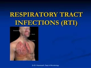
Respiratory Infections. Chapter 5. Infections of the Respiratory tract. Most common entry point for infections Upper respiratory tract nose, nasal cavity, sinuses, mouth, throat Lower respiratory tract Trachea, bronchi, bronchioles, and alveoli in the lungs. Protective Mechanisms.

Respiratory Infections
E N D
Presentation Transcript
Respiratory Infections Chapter 5
Infections of the Respiratory tract • Most common entry point for infections • Upper respiratory tract • nose, nasal cavity, sinuses, mouth, throat • Lower respiratory tract • Trachea, bronchi, bronchioles, and alveoli in the lungs
Protective Mechanisms Normal flora: Commensal organisms • Limited to the upper tract • Mostly Gram positive or anaeorbic • Microbial antagonist (competition)
Protective Mechanisms Clearance of particles and organisms from the respiratory tract Cilia and microvilli move particles up to the throat where they are swallowed. Alveolar macrophages migrate and engulf particles and bacteria in the alveoli deep in the lungs.
Other Protective Mechanisms • Nasal hair, nasal turbinates • Mucus • Involuntary responses (coughing) • Secretory IgA • Immune cells
Response towards foreign particles • Ventilatory flow • Cough • Mucociliary clearance mechanisms • Mucosal immune system
Selected Bacterial Infections Pharyngitis Group A Strep - Streptococcus pyogenes (Many viruses also cause this) Pneumonia - Streptococcus pneumoniae Diphtheria - Corynebacterium diphtheriae Tuberculosis - Mycobacterium tuberculosis Whooping cough - Bordetella pertussis
Disease of upper respiratory tract Pharyngitis and related infections Diptheria
Examples of common infections • Laryngitis • Streptococcal Pharyngitis • Scarlet Fever • Sinusitis • Diptheria • Otitis media • Advance stages – bacterial superinfection, mastoiditis, meningitis and brain abscess
Laryngitis • Most commomly upper respiratory viruses • Diphtheria - C. diphtheriae produces a cytotoxic exotoxin causing tissues necrosis at site of infection with associated acute inflammation. Membrane may narrow airway and/ or slough off (asphyxiation)
Streptococcal Pharyngitis (Strep Throat) • Caused by group A beta-hemolytic streptococci of S.pyogenes • Also causing impetigo, erysipelas and endocarditis on skin • Causing inflammation of the mucous membrane and fever; tonsilitis and otitis media may also occur • Rapid diagnosis using enzyme immunoassays.
Strep Throat • Fever • Tonsillitis • Enlarged lymph nodes • Middle-ear infection
Streptococcus pyogenes • Gram positive streptococci • Carried and transmitted from the throat • In Respiratory secretions
Group A Strep • Capsule -resistant to phagocytosis • Enzymes damage host cells • M protein adhesin The M protein has many antigenic varieties and thus, different strain of S.pyogenes cause repeat infections
Scarlet fever • Caused by S. pyogenes producing erythrogenic toxin • The bacteriophage disturb the normal characterristic of bacteria that involving genetic mutation • Also associate with pharyngitis and skin infection that caused by the same bacteria • It is nowadays become mild and rare disease
Scarlet Fever Caused by Erythrogenic Toxin secreted by S. pyogenes
Scarlet Fever • The erythrogenic toxin is coded by a gene lysogenic bacteriophage within the genome of S. pyogenes • Rash is an inflammatory reaction to the toxin
Sinusitis • Commonly caused by S. pneumoniae, Moraxella catarrhalis / H. influezae • can also caused by S. aureus/ S.pyogenes • The sinus cavity will swelling and prevent drainage that resulting pressure and severe pain • Pt usually produce mucus, bacteria and phagocytic cells that collect in the sinuses
Cont. • Chronic stages caused by Bacteriodes • Mostly occur above the root of upper teeth which infection derived from oral cavity • Treatment by applying moist heat on particular area / drop an ephedrine / raise head to help drainage • Best antibiotic - penicillin
Dipteria • Until 1935, it leading infection to children of US • Vaccine used for children is DTaP vaccine • Diptheria Toxoid antibodies Production (DTaP) • Spread thru air bourne and resistant towards drying • The greyish toungue contain fibrin, dead tissues and bacterial cells that block the air passage • 0.01mg of highly virulence toxin - fatal
Corynebacterium diphtheriae • Aerobic Gram + bacillus • Toxin inhibits protein synthesis of cells to which it binds • Destroyed cells and WBC form "pseudomembrane" which blocks airways
Corynebacterium diphtheriae • To produce toxin, C. dithpheriae must be infected with a bacteriophage carrying the toxin gene
An “AB” toxin • B = binding subunit • A = active subunit which binds to and inhibits a eucaryotic ribosomal translation factor • Vaccine is diphtheria toxoid
Otitis media • Uncomfortable common cold, that infecting nose or throat (otitis media) • 85% infecting children below 3 yrs old • Best antibiotic from penicillin group • The antibiotics used were just reduction the duration of infection
causes • S. pneumoniae • H. influenzae • S.pyogene • Moraxella catarrhalis • S.aureus
Diseases of lower respiratory tract Whooping cough Tuberculosis Pneumonia
Bordetella pertussis • Gram negative cocco-bacillus • Capsule • Adherence to ciliated cells • Pertussis toxin is A-B toxin
Pertussis (Whooping Cough) • Cough • Violent coughing followed by whooping sound • Vaccine – it is made of purified components • Not lifelong immunity – adult carriers
Bacterial Pneumonia Bacterial, viral or fungal infection can cause Inflammation of the lung with fluid filled alveoli
Community Acquired Pneumonia • Infection of the lung parenchyma in a person who is not hospitalized or living in a long-term care facility for ≥ 2 weeks • Most common pathogen = S. pneumo(60-70% of CAP cases)
“Nosocomial” Pneumonia • Hospital-acquired pneumonia (HAP) • Occurs 48 hours or more after admission, which was not incubating at the time of admission • Ventilator-associated pneumonia (VAP) • Arises more than 48-72 hours after endotracheal intubation
“Nosocomial” Pneumonia • Healthcare-associated pneumonia (HCAP) • Patients who were hospitalized in an acute care hospital for two or more days within 90 days of the infection; resided in a nursing home or LTC facility; received recent IV abx, chemotherapy, or wound care within the past 30 days of the current infection; or attended a hospital or hemodialysisclinic
Pathogenesis • Inhalation, aspiration and hematogenous spread are the 3 main mechanisms by which bacteria reaches the lungs • Primary inhalation: when organisms bypass normal respiratory defense mechanisms or when the Pt inhales aerobic GN organisms that colonize the upper respiratory tract or respiratory support equipment
Pathogenesis • Aspiration: occurs when the Pt aspirates colonized upper respiratory tract secretions • Stomach: reservoir of GNR that can ascend, colonizing the respiratory tract. • Hematogenous: originate from a distant source and reach the lungs via the blood stream.
Pathogens • CAP usually caused by a single organism • Even with extensive diagnostic testing, most investigators cannot identify a specific etiology for CAP in ≥ 50% of patients. • In those identified, S. pneumo is causative pathogen 60-70% of the time
Streptococcus pneumonia • Most common cause of CAP • Gram positive diplococci • “Typical” symptoms (e.g. malaise, shaking chills, fever, rusty sputum, pleuritic hest pain, cough) • Lobar infiltrate on CXR • Suppressed host • 25% bacteremic
Pneumonia Atypical Pneumonia • #2 cause (especially in younger population) • Commonly associated with milder Sx’s: subacute onset, non-productive cough, no focal infiltrate on CXR • Mycoplasma: younger Pts, extra-pulm Sx’s (anemia, rashes), headache, sore throat • Chlamydia: year round, URI Sx, sore throat • Legionella: higher mortality rate, water-borne outbreaks, hyponatremia, diarrhea
Viral Pneumonia • More common cause in children • RSV, influenza, parainfluenza • Influenza most important viral cause in adults, especially during winter months • Post-influenza pneumonia (secondary bacterial infection) • S. pneumo, Staph aureus
Other bacteria • Anaerobes • Aspiration-prone Pt, putrid sputum, dental disease • Gram negative • Klebsiella - alcoholics • Branhamella catarrhalis - sinus disease, otitis, COPD • H. influenza • Staphylococcus aureus • IVDU, skin disease, foreign bodies (catheters, prosthetic joints) prior viral pneumonia
Streptococcus pneumoniae • Pneumococcus • Encapsulated • Often secondary infection following influenza virus
Bacterial Pneumonia • Streptococcus pneumoniae • 2/3 of all pneumonia • Risk Factors- old age, season, underlying • viral infection, diabetes, alcohol and narcotic use • Variable capsular antigen • Purified component (capsule) vaccine • Others that cause pneumonia: • Mycoplasma pneumoniae • Legionella pneumophila
Mycobacterium tuberculosis • Acid-fast bacillus – complex cell wall with “cord factor” • Causes TB: lungs bones, other organs • Airborne, (milk, v. rare)
Mycobacterium tuberculosis • Thick lipid coat of “Mycolic fatty acids” • Grows very slowly • Resists killing by macrophages and grows in them
Tubercule formation A tubercle in the lung is a “granuloma” consisting of a central core of TB bacteria inside an enlarged macrophage, and an outer wall of fibroblasts, lymphocytes, and neutrophils
Tuberculosis • Primary • Lung tubercles, caseous, tuberculin skin reaction • Secondary (reactivation) • Consumption: Coughing and chronic weight loss • Dissemination • Extrapulmonary TB (lymph nodes, kidneys, bones, genital tract, brain, meninges)