1 / 1

NEN_72_12_2013_10_08_HAMADA_JNEN13-115_SDC4

sydnee-case
Télécharger la présentation

NEN_72_12_2013_10_08_HAMADA_JNEN13-115_SDC4

An Image/Link below is provided (as is) to download presentation Download Policy: Content on the Website is provided to you AS IS for your information and personal use and may not be sold / licensed / shared on other websites without getting consent from its author. Content is provided to you AS IS for your information and personal use only. Download presentation by click this link. While downloading, if for some reason you are not able to download a presentation, the publisher may have deleted the file from their server. During download, if you can't get a presentation, the file might be deleted by the publisher.

E N D

Presentation Transcript


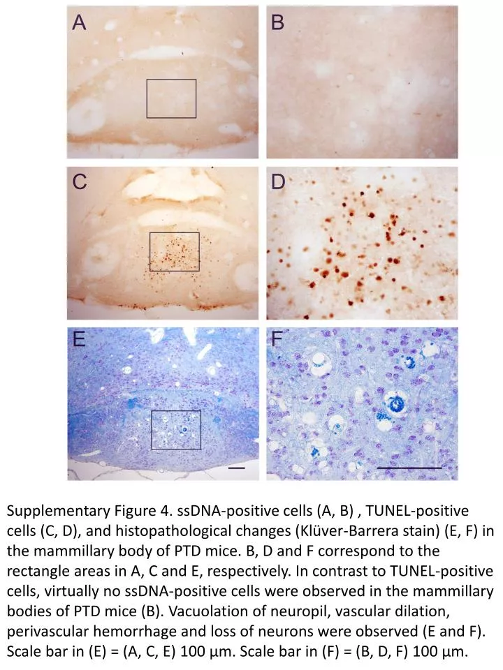
  1. Supplementary Figure 4. ssDNA-positive cells (A, B) , TUNEL-positive cells (C, D), and histopathological changes (Klüver-Barrera stain) (E, F) in the mammillary body of PTD mice. B, D and F correspond to the rectangle areas in A, C and E, respectively. In contrast to TUNEL-positive cells, virtually no ssDNA-positive cells were observed in the mammillary bodies of PTD mice (B). Vacuolation of neuropil, vascular dilation, perivascular hemorrhage and loss of neurons were observed (E and F). Scale bar in (E) = (A, C, E) 100 μm. Scale bar in (F) = (B, D, F) 100 μm.

More Related