Download
1 / 32
320 likes | 508 Vues
Mediastinal Masses 2010 WOFAPS Meeting. George W. Holcomb, III, M.D., MBA Surgeon-in-Chief Children’s Mercy Hospital Kansas City, Missouri. Diseases in the Mediastinum. Infection Tumors – Benign/Malignant Lympadenopathy Cysts Lymphangioma. Symptoms. Severe Respiratory Distress.

E N D
Mediastinal Masses2010 WOFAPS Meeting George W. Holcomb, III, M.D., MBA Surgeon-in-Chief Children’s Mercy Hospital Kansas City, Missouri
Diseases in the Mediastinum • Infection • Tumors – Benign/Malignant • Lympadenopathy • Cysts • Lymphangioma
Symptoms Severe Respiratory Distress Asymptomatic
Regionalization > Teratoma, Thymus Ectopic Thyroid Adenopathy Adenopathy Bronchogenic Cysts Esophageal Duplication Cysts Neurogenic Tumors Esophageal Duplication Cysts Anterior Mediastinum Middle Mediastinum Posterior Mediastinum > >
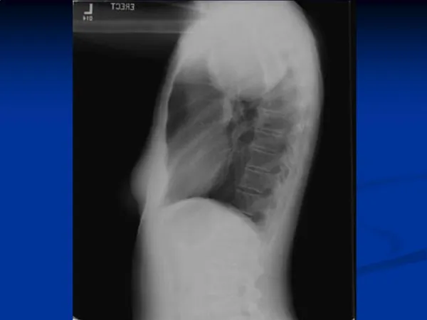
Imaging Studies • CT (IV, Oral contrast) • MRI (esp. posterior mediastinal lesion) • Contrast esophagram • Technetium – 99m pertechnetate scan
Imaging Studies • Contrast esophagram
Anterior Mediastinum • Lymphoma (50% of all mediastinal lesions) • Teratoma • Germ cell tumors • Cystic hygromas • Thymic lesions
Anterior MediastinumLymphoma • Usually older child • Hodgkin’s – 14 yrs • Non-Hodgkin’s – 9 yrs • Often have other symptoms and other adenopathy • Frequently have airway compromise
Anterior MediastinumAirway Compromise • Tracheal collapse on induction of anesthesia • Look diligently for extrathoracic approach & local anesthesia • Cervical adenopathy • Pleural effusion • Bone marrow examination • Needle biopsy often obtains inadequate tissue, esp. in Hodgkin’s • ? Short course steroids or radiation followed by thoracoscopic biopsy or mini-thoracotomy
Middle Mediastinum • < 2 yrs: Remnants of embryonic foregut (trachea & esophagus) • Pericardial cysts • Lymphadenopathy
Middle Mediastinum Bronchogenic Cyst • Can be adjacent to or far away from bronchogenic structures • Usually have respiratory epithelium • Can be large and cause respiratory symptoms Esophageal Duplication Cyst • Adjacent to or embedded in wall of esophagus • Can have respiratory or GI epithelium • May either obstruct or erode through esophageal wall • Thoraco-abdominal duplications: orginate near duodenum & jejunum and expand to middle mediastinum (gastric mucosa & vertebral anomalies)
ThoracoscopyTechnique • Baseball diamond concept for location of instrument sites • Convert if difficult
Thoracoscopy Single lung ventilation
Posterior Mediastinum • Ganglioneuroma • Ganglioneuroblastoma • Neuroblastoma
Posterior MediastinumNeuroblastoma • Very good prognosis, especially Stage I & II *Paraplegia implies compression of spinal cord (MRI & urgent laminectomy)
Thoracoscopic Operations Children’s Mercy Hospital (2000-2007) IPEG 2007 J LAST 18:131-135, 2008
ThoracoscopyPearls & Pitfalls • Single lung ventilation, if possible • Keep dissection as close to the wall of a foregut duplication cyst to avoid entry into an adherent structure (esophagus, bronchus) • Aspirate large cystic mass, if necessary • Place bougie in esophagus to identify its location • If common wall of duplication left intact, it is imperative to remove mucosal lining