Methods for Gene Activity Analysis
Methods for Gene Activity Analysis. By Auni Hovanesian Krista Templeton. What Methods Can We Use to Study Gene Expression?. Three Basic Approaches: RT-PCR GeneChip Microarray Upstream Regulatory Region Analysis. What Is The Basis of RT-PCR Activity Analysis?. mRNA!.


Methods for Gene Activity Analysis
E N D
Presentation Transcript
Methods for Gene Activity Analysis By Auni Hovanesian Krista Templeton
What Methods Can We Use to Study Gene Expression? • Three Basic Approaches: • RT-PCR • GeneChip Microarray • Upstream Regulatory Region Analysis
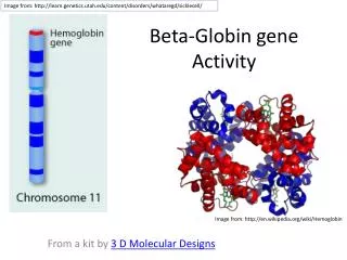
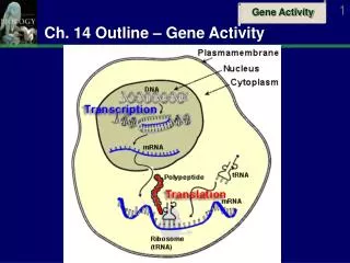
What Is The Basis of RT-PCR Activity Analysis? mRNA! • mRNA is only present in cells for genes that have been transcribed and will usually be actively expressed once translated into proteins • RT-PCR links gene expression to mRNA presence in different compartments of plant • But in order to trace mRNA back to an original gene, must amplify sample, since often only small amounts present
Why RT-PCR Rather Than Regular PCR? • mRNA will not work in our PCR reaction, so need to convert it to cDNA • reagents being used in PCR are DNA-specific (i.e. DNA polymerase) • DNA is more stable than RNA • More practical for long-term storage purposes • RNA’s instability would give lower yield in PCR
AAAAAAA So How Does RT-PCR Work? • Isolate mRNA from area-specific plant tissue/organ samples • Convert all isolated mRNA strands to cDNA using Reverse Transcriptase mRNA Reverse Transcriptase cDNA
3) Use first cDNA template as now compatible basis of PCR: • Only need to synthesize a single strand of cDNA template to start PCR • One cDNA representing every mRNA in each sample--like gene expression library • PCR is like selection of a particular gene from the library (if it is there!) • Gene-specific primers designed in exons (due mRNA splicing) • If PCR product formed with gene-specific primers for RT-PCR, will have amplified the cDNA correlate of original gene • Indicates presence of mRNA correlating to gene of interest • Helps localize gene activity to wherever sample came from
Sample RT-PCR Results 100 bp Silique RT+ Positive Control Negative Control Silique RT- Leaf RT+ Leaf RT- Tubulin Bands 475 bp Hypothetical cDNA Band 200 bp
So How Do We Verify and Further Specify RT-PCR Results? GeneChip Microarray Analysis!
What is a GeneChip Microarray? • Using cDNA created from the mRNA isolated from various organs, we can analyze the mRNA accumulation levels for all genes • Done by creating the complementary strands of all the known gene sequences and assembling them on a chip • Different chips are used for various stages of development • The cDNA sequences are tagged with flourescent labels that glow a certain color when in contact with the complementary strand; colors read by a computer
What is the Resulting Image? Red means active Green means not as active No color means no activity
So, How Do RT-PCR and GeneChips Complement Each Other? • RT-PCR may be slightly more accurate, since working with smaller fragments (only portions of cDNA) • But GeneChips provide more specificity more efficiently • The two combined provide convenient checkpoints for each other’s results • both working with same principle of mRNA level analysis to determine gene expression
Overview • The upstream regulatory region of a gene contains its “on” switch. • Once the upstream region is fused to GFP (green fluorescent protein) or GUS (betaglucuronidase) it can be transformed into the Arabidopsis plant • Once transformation has occurred gene expression will indicate where the gene of interest is transcribed.
Strategy of Promoter Activity Analysis • Arabidopsis Genomic DNA • PCR amplification of upstream region • With Gene-specific Primers • And High Fidelity DNA Polymerase PCR Product pENTR/D-TOPO vector Ligation: Population of Recombinant Plasmid (vector+PCR product) and NON-recombinant plasmid (vectory only) Transformation of competent E.coli cells Screening for E.coli cells harboring recombinant plasmid Confirmed Recombinant plasmid DNA: Verifying the authenticity of recombinant plasmid DNA by Restriction Enzyme Digestion Recombinant Plasmid DNA + Beta-Gluronidase (GUS) gene carrying T-DNA Vector DNA sequences: verification of the cloned Promoter Region by Sequencing Analysis. Sequence Analysis and confirmed identity of the cloned upstream region
What Are the Steps Required to Isolate the Promoter Region? • Conduct PCR on the Arabidopsis DNA in order to amplify the upstream region with iProof polymerase
Why Use a Proofreading Polymerase? • iProof polymerase corrects nucleotide errors during amplification • One mutation could affect the transcription of a gene
What To Do After the Promoter Region Has Been Ampified • The section needs to be enzymatically inserted into a plasmid vector where it can be placed into an E.coli cell. DNA pENTR-TOPO vector Recombinant Plasmid Mix E.coli cells with plasmids in presence of CaCl. Culture on nutrient agar plates containing ampicillin.
What Does Topoisomerase 1 Do? • The pENTR/d-TOPO vector contains Topoisomerase 1 • Relieves supercoils in circular DNA plasmids by nicking one of the strands of the DNA double helix • Linearized the pENTR vector, allowing insertion of the PCR fragment • Re-ligates vector
How Do GUS and GFP Work and What Are Their Differences? • A mature Arabidopsis embryo expressing Green Fluorescent Protein GUS is the more sensitive of the two Common Reporter Genes • GUS and GFP connect to the promoter region. When the genes are turned on through the promoter region GUS and GFP are turned on also
Acknowledgements • Jordan, Jennifer, and Brian from Spring 2006 for some useful slide ideas and diagrams • Kelli for her presentation on Upstream Region Analysis • Brandon for all his tech support! • Anhthu, Bekah, and Daisy for their help and many explanations