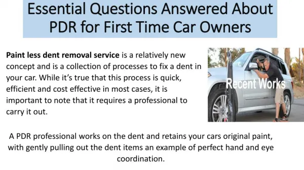
Download

1 / 32
320 likes | 551 Vues
DENT 5315/DH 2215 February 8, 2008. Dr. Sandra Myers myers025@umn.edu. What are elephant tusks made of?. Enamel Dentin Enamel & Dentin Chalk Marble. Ectoderm & Enamel. Enamel: an epithelially derived protective covering for the teeth derived from ectoderm Fig. 2-12 p. 26.

E N D
DENT 5315/DH 2215 February 8, 2008 Dr. Sandra Myers myers025@umn.edu
What are elephant tusks made of? Enamel Dentin Enamel & Dentin Chalk Marble
Ectoderm & Enamel Enamel:an epithelially derived protective covering for the teeth derived from ectoderm Fig. 2-12 p. 26
Ectoderm & Enamel What happens when ectoderm fails to form or form properly?
Enamel & Amelogenesis Enamel: most highly mineralized extracellular matrix 96% mineral 4% organic material & water
Enamel & Amelogenesis Hard Tissue Formation Amelogenesis Structure of Enamel Clinical Correlations
Hard Tissue Formation “Bell Stage” Hard Tissue Formation Amelogenesis Structure of Enamel Clinical Correlations
Amelogenesis 3 Main Functional Stages: 1. Presecretory 2. Secretory 3. Maturation Presecretory Ameloblasts: Differentiate (acquire phenotype) Change polarity (nuclei) Develop enamel synthesis apparatus Morphodifferentiation (shape) Histodifferentiation (microscopic)
Amelogenesis Begins first at cusp tips Then sweeps down crown slopes Stops at CEJ
Amelogenesis - Secretory Stage pcw = proximal cell web dcw= distal cell web cell webs hold cells in formation
Amelogenesis - Secretory Stage Hallmarks: Intense synthetic & secretory activity Secretion is continuous Secretory granules not stored Almost immediate mineralization Initial layer does not contain rods Enamel Matrix: Note Tomes’ processes & picket-fence appearance.
Amelogenesis - Secretory Stage IGS = interrod, RGS = rod growth sites sg = secretory granules, ppTP = proximal dp = distal portion of Tomes process
Amelogenesis - Secretory Stage Enamel crystals: What are these composed of? Initial enamel: “no rods” crystalline calcium phosphate (hydroxyapatite) substituted with carbonate ions “pits filling with enamel”
Enamel - Amelogenesis (Note how trajectory of enamel rods changes)
Amelogenesis - Life Cycle of Ameloblasts Functional stages in life cycle of ameloblasts: Morphodifferentiation Histodifferentiation 3. Secretory (initial) 4. Secretory (Tomes’ process) Maturation (ruffle-ended) Maturation (smooth-ended) Protective
Amelogenesis - Maturation Stage Maturation Process: Removal of water & organic material Introduction of additional inorganic material Process = “Modulation” cyclic creation, loss, and recreation of highly invaginated ruffle-ended apical surface on ameloblasts
Smooth-ended Ruffle-ended Amelogenesis - Maturation Stage Ameloblasts incorporation of inorganic material exit of protein fragments & water Ameloblasts
Ameloblast Modulation (a visually dramatic activity) regional pH variations maturing enamel (rat incisors) Large bands = ruffle-ended cells Smaller bands = smooth-ended cells
Amelogenesis - Maturation Stage Enamel hardens before tooth erupts Results from growth in width, thickness of crystals Amelogenesis slow process almost mature enamel, most mineral removed
Amelogenesis - Enamel Proteins Enamel Proteins: (Table 7-2 text) 1. Contributing to appositional growth, thickness enamel * Amelogenin (main protein in forming enamel) * Ameloblastin * Enamelin 2. Postsecretory processing & protein degradation 3. Related to basal lamina covering maturing, preeruptive enamel 4. Legacy proteins
Amelogenesis - Enamel Proteins Amelogenin protein (stained red) Ameloblasts http://dentistry.uic.edu/CraniofacialGenetics/ResearchTED.htm
Amelogenesis - Enamel Proteins Amelogenin vs Ameloblastin
Protective Stage Full thickness of enamel complete, enamel mature Ameloblast layer & papillary layer form “reduced enamel epithelium” What is the enamel space?
Enamel - Structure Hard Tissue Formation Amelogenesis Structure of Enamel Clinical Correlations Scanning Electron Microscopy R = Rod & IR = Interrod Areas
Enamel - Structure Aapd.org/publications/peddent/ Note: rod, interrod crystals same, but divergent orientation
Enamel - Structure Enamel: hydroxyapatite crystals Young Enamel Older Enamel Transmission EM: rod surrounded by interrod enamel
hexagonal contour to older mature crystals recently formed thin crystals Enamel - Structure Crystals
Enamel - Structure Crystal Profiles hexagons with unequal-sided peaks (un) & equal-sided peaks (eq) x 300,000 (rat incisor)
Enamel - Structure enamel rod orientation A: alternating orientations B: row arrangement C: note thin, long apatite crystals
Enamel - Structure rod sheath rod sheath = boundary between rod & interrod enamel, contains organic material Cat Secretory Stage Enamel Mature Cat Enamel
Enamel - Structure 3 Faces of an Enamel Block cross-section of rod-interrod area appearance compared to “keyhole”