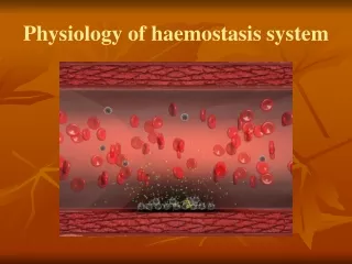
Haemostasis
Haemostasis. Tiffany Shaw MBChB II 2002. Haemostasis Pathway. Injury Collagen exposure Tissue Factor Platelet adhesion Coagulation Cascade Release reaction

Haemostasis
E N D
Presentation Transcript
Haemostasis Tiffany Shaw MBChB II 2002
Haemostasis Pathway Injury Collagen exposure Tissue Factor Platelet adhesion Coagulation Cascade Release reaction Platelet aggregation Fibrin Primary haemostatic plug Secondary haemostatic plug
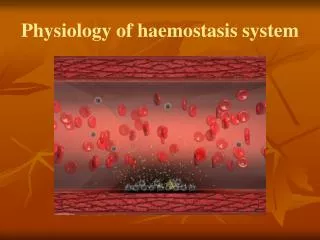
Integrity of Haemostasis Depends on: • Vessel wall • Platelets (no. and function) • Coagulation factors
Vessel Wall • Release tissue factor that initiates coagulation cascade. • Exposed collagen results in platelet adhesion (using vWF as a bridge). • Hereditary haemorrhagic telangiectasia • Scurvy • Senile purpura
Low Platelet Number • N = 150 – 450 • Excessive bleed / Spontaneous bleed • Mucosal surfaces / Skin • Immune Mediated Thrombocytopenia • Drug induced • Haemolytic Uraemic Syndrome • DIC
Platelet Dysfunction • Abnormal platelet aggregation • Normal platelet count • Excessive / spontaneous bleed • Mucosa / Skin • Inherited disorders • Drug induced
Coagulation Factors Deficiency • Spontaneous bleeding into deep tissues • Increased post-traumatic haemorrhage • Factor 8 – Haemophilia A • Factor 9 – Haemophilia B • vWF – Von Willebrand’s disease Von Willebrand’s Factor: • Protein produced by endothelial cells which mediates platelet adhesion to endothelium and carries factor 8 in plasma.
Haemaphilia A • Factor 8 deficiency • 50 / million people • Sex-linked • Most detected in childhood • Mild symptoms ~ spontaneous bleed • Chronic joint disease after repeated bleed
Haemophilia B • Factor 9 deficiency • = Christmas disease • Also sex-linked • Similar presentation as haemophilia A • Much rare
Von Willebrand’s Disease • vWF deficiency • Autosomal dominant (F = M) • Most common inherited bleeding disorder • Bleeding from mucosa • Excessive blood loss in trauma • Bleeding into deep tissue (rarer)
Liver Disease • Mixed – both coag deficiency and platelet problem • Reduced coag factor synthesis (1, 2, 5, 7, 9, 10) • Cirrhosis Hypersplenism Thrombocytopenia
DIC Generalised activation of coag cascade Wide-spread fibrin formation Thrombosis Low coag factor Bleeding
DIC Consequence of Thrombosis: • Tissue infarction Renal impairment • CVA • PE • DVT Consequence of Bleeding: • Purpura • GI Bleed
DIC Causes of coag cascade activation: • Sepsis • Disseminated cancer • Obstetric complications (e.g. retained product of conception) • Fulminant liver disease • Anaphylaxis • AML
Drug Induced Coag Factor Def Heparin: • Inactivates certain coag factors (e.g. 9a, 10a, 11a, 2) • Also impairs platelet function Warfarin: • Block synthesis of Vit K dependent factors (2, 7, 9, 10)
Other Disorders • Vitamin K deficiency • Autoantibodies to coag factors • Haemorrhagic disease of the newborn • Massive transfusion
Investigations for Bleeding • Platelet Count • Bleeding time • Activated Partial Thromboplastin Time • Prothrombin Time (INR) • Plasma level assay
Management • Replace deficiency • Treat underlying cause • Reversal of drug effects • Supportive therapy
Extra Notes on ITP • Platelets become coated by autoantibodies • Removed by RES reduced lifespan • Low platelet count • Acute / Chronic • Purpura / Spontaneous bleed / Asymptomatic • May resolve spontaneously or require medical treatment
Extra Notes on ITP • Isolated low platelet count < 20 • Increased megakaryocytes in bone marrow • Normal INR + APPT • Increased bleeding time
Extra Notes on ITP Mx: • Do nothing • Prednisolone • Splenectomy • Other immunosuppresives
Extra Notes on IPT May be associated with: • Haematological malignancies • Viral infections (e.g. HIV) • C.T. disorders (e.g. SLE) Test for c.t. disorders.
70-Year-Old with ITP • Incidental finding in 1993 • Platelet count = 60 • Asymptomatic • Referred to haematologist • Test for c.t. disorders • U/S for spleen size • Bone marrow biopsy
70-Year-Old with ITP • Negative for c.t. disorders • Spleen size normal • Bone marrow: increased megakaryoccytes • Continue monitoring (weekly FBC)
70-Year-Old with ITP • December 1993 • Bruising with minimal trauma • Platelet count = 43 • Start Prednisolone • After 6/52 platelet count 190 • Slowly dropped again after withdrawal • Asymptomatic
70-Year-Old with ITP • Continue monitoring • Platelet transfusion if serious bleeding occurs • Consider splenectomy if worsens • Discharged to GP last week