Download
1 / 1
10 likes | 146 Vues
THE EFFECT OF HIGH SALT CONCENTRATION ON PROTEIN DYNAMICS: MOLECULAR DYNAMICS SIMULATIONS OF P.WOESEI TATA BOX-BINDING PROTEIN. Nina Pastor 1 and Harel Weinstein 2

E N D
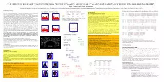
THE EFFECT OF HIGH SALT CONCENTRATION ON PROTEIN DYNAMICS: MOLECULAR DYNAMICS SIMULATIONS OF P.WOESEI TATA BOX-BINDING PROTEIN. Nina Pastor1 and Harel Weinstein2 1Facultad de Ciencias, UAEM, Av. Universidad 1001, Col. Chamilpa, 62210 Cuernavaca, Morelos, México and Dept. of Physiology and Biophysics, 2Mount Sinai School of Medicine, One Gustave L. Levy Place, New York, NY 10029, U.S.A.. • SUMMARY OF H-BOND AND WATER BRIDGE INTERACTIONS • ATH2 ( 19) 1 SGIVPTLQNI VSTVNLDCKL DLKAIALQAR NAEYNPKRFA AVIMRIREPK • SCE ( 61) 1 SGIVPTLQNI VATVTLGCRL DLKTVALHAR NAEYNPKRFA AVIMRIREPK • PWO ( 5) 1 SKVKLRIENI VASVDLFAQL DLEKVLDLCP NSKYNPEEFP GIICHLDDPK • |------------| |-----| |-| |---| • S1 H1 S2 S3 • ATH2 51 TTALIFASGK MVCTGAKSED FSKMAARKYA RIVQKLGF-PA KFKDFKIQNI • SCE 51 TTALIFASGK MVVTGAKSEDDSKLASRKYA RIIQKIGF-AA KFTDFKIQNI • PWO 51 VALLIFSSGK LVVTGAKSVQ DIERAVAKLA QKLKSIGVKFK RAPQIDVQNM • |---| |------| |-----------------| |------ • S4 S5 H2 • ATH2 101 VGSCDVKFPI RLEGLAYSHA AFSSYEPELF PGLIYRMKVP KIVLLIFVSG • SCE 101 VGSCDVKFPI RLEGLAFSHG TFSSYEPELF PGLIYRMVKP KIVLLIFVSG • PWO 102 VFSGDIGREF NLDVVALTLP N-CEYEPEQF PGVIYRVKEP KSVILLFSSG • -------| |-----||-| |---| |---| • S1’ H1’ S2’ S3’ S4’ • ATH2 151 KIVITGAKMRDETYKAFENI YPVLSEFRKI • SCE 151 KIVLTGAKQREEIYQAFEAI YPVLSEFRKM • PWO 151 KIVCSGAKSE ADAWEAVRKL LRELDKYGLL • |-----| |-----------------| • S5’ H2’ • Secondary structure assignment shown below the sequences. • Red: water bridges; teal: water bridges induced in salt; pink: water bridges lost in salt. • Underlined: sc – sc and sc – mc H-bonds. Wavy underline: H-bonds induced in salt. Double underline: H-bonds lost in salt. • INTRODUCTION • The TATA box-binding protein (TBP) is a general transcription factor present in archaea and eukaryotes; it is required for transcription by the three nuclear RNA polymerases. • TBP is conserved throughout evolution, both in structure and in function. In transcription mediated by RNA polymerase II, TBP binds to the TATA box, and this binary complex recruits TFIIB. The structure of this ternary complex is also conserved throughout evolution. • The structures of the C-terminal domain of TBPs from plants (A.thaliana – ATH [1]), yeast (S.cerevisiae – SCE [2]) and a hyperthermophile (P.woesei – PWO [3]) have been solved by X-ray crystallography, revealing practically the same structure for all, as expected from the ~40% sequence identity between these proteins. A distinguishing feature of PWO TBP is a disulfide bond, that is absent in the other two TBPs. • TBP molecules in different organisms are subject to a wide variety of environments. ATH and SCE represent mesophile organisms; the optimal growth temperature for yeast is ~30C. On the other hand, PWO grows optimally at 105C, with an internal salt concentration of 0.8 M. The thermal stability of TBPs from different organisms varies widely, and depends also on the salt concentration: SCE will denature at 60C in 50 mM salt, but it will denature even at room temperature in salt concentrations over 200 mM. PWO TBP denatures at 101C in 50 mM salt, but at 109C in 800 mM salt. Under reducing conditions, the melting temperature decreases to 97C [3]. • The thermal stability of PWO TBP has been rationalized [3] from an analysis of the crystal structure on the basis of the unique disulfide bond, an increase in ion pairs on the surface of TBP, an increase in buried surface area, and a more compact packing. The halostability can be rationalized on the basis of an increase in the number of acidic residues, compared to the mesophilic TBPs. • Given the phylogenetic conservation in function of TBP, it is expected that • structure and dynamics that relate to function will be common to ATH, SCE and PWO TBPs. • structural and dynamic features that are specific to PWO TBP will indicate adaptation mechanisms to extreme environments. • APPROACH: • We carried out MD simulations of ATH, SCE and PWO monomers, at 300K, in TIP3 water, for 2 ns, with the CHARMM23 program and potential. PWO was also simulated with TIP3 water and 0.75 M NaCl (PWOSAL). BACKBONE FLUCTUATIONS SIDE CHAIN DYNAMICS: ATH SCE PWO PWOSAL sc entropy 175±9 166±3 180±7 188±3 sc TS 52±3 50±1 54±2 56±1 Entropy in cal/molK from S = -R∑pilnpi, where pi is the probability of populating a particular rotamer; TS calculated at 300K, and given in Kcal/mol. • FINDINGS 2: • H-BONDS AND HYDRATION • The relative sizes of polar (46%) and apolar (54%) surfaces in PWO are slightly different for mesophile TBPs (42% polar and 58% apolar). • The increase in polar solvent accessible area in PWO relative to ATH and SCE, is also reflected in a greater number of H-bonds to water during the simulations. • More water molecules are coordinated by PWO due to the larger number of D and E residues in PWO compared to ATH and SCE. D residues are ~20% better at forming water bridges than E residues. • There is a common water bridge network in all the simulations, linking the C-terminus to helix 1’ and to the stirrup below. • PWO is more densely packed than the mesophile TBPs (1.4% smaller volume). • The amount of H-bonds between side chains is larger in PWO and PWOSAL than in ATH and SCE. • PWO and PWOSAL have more and larger networks of H-bonded residues, bridging elements of secondary structure and probably conferring structural stability. • Mesophile TBPs tend to have more H-bonds in the N-terminal subdomain than in the C-terminal subdomain. This pattern is reversed in PWO and PWOSAL, probably due to the presence of the disulfide bond in the N-terminal subdomain. H-BOND NETWORKS: ATH: K151 - D105 - K107 (S5’) (S1’) (S1’) loop S3-S4 | K78 - Y79 - T51 (H2) (H2) (S4) • EFFECTS OF THE ADDITION OF SALT • Increase in solvent accessible area, but not in a change of the relative amounts of polar (46%) and apolar (54%) surfaces. • No change in the number of coordinated water molecules, but an increase the number of water bridges between the side chains in PWOSAL. • Slight increase in molecular volume, suggesting that salt makes the protein swell. • Swelling and increase in backbone fluctuations also evident from the decrease in the number of H-bonds between main chain atoms in PWOSAL compared to PWO. Some of the regions with increased backbone fluctuations correspond to the places where mc-mc H-bonds were either debilitated or lost. Overall decrease in the number of direct H-bonds in PWO. • No change in the number of H-bonds between side chains. Nevertheless, there is a redistribution of the H-bonds. The actual H-bond networks are different in the two simulations, and are spread over larger areas in PWOSAL. This is probably due to the fact that the internal water molecule in PWO tried to escape to the bulk solution, causing an increase in the distance between strands 1’ and 5’ (see also the stirrup – stirrup distance distribution), and disrupting the network underlining the bottom face of PWO. SCE: loop S3-S4 | K78 - Y79 - T51 | R45 - E33 (S3) (S2) • CONCLUSIONS • Structure stabilization in TBPs • The N-terminal subdomain seems to be the weakest part of the structure. In mesophiles it is stabilized by small networks of H-bonds and water bridges (linking, for example, helix 2 to strand 4), while in PWO it is stabilized by a disulfide bond linking H1 to the body of the protein. • The stabilization of the C-terminal subdomain is accomplished through both water and H-bond networks. These are larger and more complex in PWO than in the mesophiles. • The addition of salt results in a “swelling” of the PWO structure, with the loss of some main chain H-bonds, but with a gain of water bridges between side chains (especially D). Halophilicity in this protein is reflected in better hydration, mediated principally by the larger number of D residues, but not by specific binding of ions. • The increase in H-bond and water bridge formation does not necessarily lead to an entropic penalty. The H-bonds and water bridges are under constant formation and breaking, thus conferring an enthalpic and an entropic advantage to PWO and PWOSAL. • Functional inferences • TBP does not collapse in the absence of DNA, as seen in a previous simulation [4]. • The two subdomains display twisting and compression-extension type motions, a flexibility which is probably related to the ability of TBP to bind DNA sequences with different degrees of bendability. • The addition of salt changes the dynamics of PWO such as to bring it closer to the behavior of the mesophilic TBPs, in agreement with the “corresponding states” hypothesis [5]. PWO: K151 - D106 - R109 | C-ter (H2’) (S3’) E173 - Y135 || (S2’) (H1’) R136 - E124 = C123 - A117 || Y125 - D114 (loop S1’-H1’) • FINDINGS 1: • OVERALL DYNAMICS • Backbone atomic fluctuations reveal similarities and individual differences in the dynamic patterns of the protein. The highest mobility regions correspond to the stirrups (residues 30-40 and 125-135, approximately), and also to the loop connecting helix 2 to strand 1’ (see C trace and alignment). • Global motions of the protein, monitored by the stirrup – stirrup distance, and the distances and angles between helices 1 and 1’, and 2 and 2’, show no evidence of collapse of the protein in any of the simulations. PROTEIN-PROTEIN HYDROGEN BONDS: ATH SCE PWO PWOSAL sc-sc 19.8 24.2 26.6 26.7 mc-mc 172.2 173.7 179.7 170.7 mc-sc 19.7 14.5 12.9 11.8 total 211.7 212.4 219.2 209.2 PROTEIN-WATER HYDROGEN BONDS: ATH SCE PWO PWOSAL Hbonds to water 387.3 398.1 441.2 444.1 sc-sc water bridges 46.0 49.5 63.8 67.1 sc-mc water bridges 29.3 27.5 33.5 33.3 Total water bridges 75.2 77.0 97.3 100.4 • EFFECTS OF THE ADDITION OF SALT • The dynamic behavior: PWO becomes closer to the mesophile TBPs, except for the H1 – H1’ distance distribution. • Increase in backbone fluctuations: general increase, except at the N-terminus and the C-caps of helices 2 and 2’ (c.f. PWO and PWOSAL). The most affected regions correspond to surface exposed loops, a region in the middle of helix 2 (A80-L83), and strand 4’ (S148-K151). • Side chain entropy: (estimated by monitoring the population of the different rotamers during the simulations) an apparent increase in side chain entropy, mainly of the hydrophobic residues. Most of the charged amino acids experience a decrease in conformational entropy. • Salt condensation: As expected for a macromolecule with a total charge of +1e, PWOSAL shows no condensation. 18% of Na and Cl atoms were found within 5.5 Å of the surface of PWOSAL, and no particular association lasted more than 250 ps, suggesting that salt stabilization is not due to specific binding to the TBP. PWOSAL: (S5’) (S1’) K151 - D106 - R109 S155 - N100 | || C-ter Q99 - S13 (S1) (H2’) (S3’) (H2’) (H2’) (H2’) E173 - Y135 - K169 - E165 - R168 || (S2’) R136 - E124 (S1) (S5) (loop S4-S5) D15 - K60 - S58 REFERENCES: [1] Nikolov, D.B. and Burley, S.K. (1994) Nat. Struct. Biol. 1:621-37. [2] Chasman, D.I., et al. (1993) Proc. Natl. Acad. Sci. 90:8174-8. [3] DeDecker, B. S., et al. (1996) J. Mol. Biol. 264:1072-84. [4] Miaskiewicz, K. and Ornstein, R.L. (1996) J.Biomol. Struct. Dyn. 13:593 [5]Jaenicke, R. and Bohm, G. (1998) Curr. Opin. Struct. Biol. 8:738-48.