Download
1 / 34
380 likes | 901 Vues
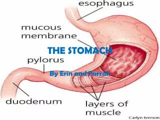
INFLAMMATIONS OF THE STOMACH CHRONIC AUTOIMMUNE GASTRITIS. Extensive multifocal atrophy (atrophic gastritis) Endemic in some parts of the world, e.g. Japan Pathogenesis: Autoantibodies to gastric glands parietal cells Gland destruction & mucosal atrophy

E N D
INFLAMMATIONS OF THE STOMACHCHRONIC AUTOIMMUNE GASTRITIS • Extensive multifocal atrophy (atrophic gastritis) • Endemic in some parts of the world, e.g. Japan • Pathogenesis: • Autoantibodies to gastric glands parietal cells • Gland destruction & mucosal atrophy • Loss of acid & IF production (pernicious anemia) • Pathology: variable gland loss, atrophy & intestinal metaplasia; dysplasia of metaplastic epithelium • If severe parietal cell loss: hypo- or achlorhydria & hypergastrinemia • Px: 2 - 4% risk of developing gastric carcinoma
PATHOLOGY OF THE STOMACHHYPEPTROPHIC GASTRITIS • Group of uncommon conditions characterized by enlargement of rugal folds of gastric mucosa • Three main variants: • Menetrier’s disease: rare idiopathic disease; may be asymptomatic or produces pain, nausea, vomiting & bleeding; protein-losing gastroenteropathy • Hypersecretory gastropathy: associated with hyperplasia of parietal and chief cells • Gastric gland hyperplasia: secondary to excessive gastrin secretion by a gastrinoma (Zollinger-Ellison syndrome) • Radiologically or endoscopically may mimic carcinoma
PATHOLOGY OF THE STOMACHGASTRIC EROSIONS & ULCERATIONS • Erosion: loss of superficial epithelium of mucosa • May heal within days • Ulcer: breach in the mucosa, which extends through the muscularis mucosa into the submucosa or deeper • Needs longer time to heal • Main types: • Acute gastric erosions & ulcerations • Peptic ulcers
GASTRIC ULCERATIONSPEPTIC ULCER • Chronic, most often solitary, lesions that occur in any part of GIT exposed to aggressive action of acid-peptic juices • Sites: 98% occur in either the duodenum or stomach (4:1 ratio) • Patients: common in industrialized countries: 1-2% of population have active disease; autopsy studies: 6-14% for men, 2-6% women • Remitting-relapsing lesions, mostly in middle-aged to older adults • Pathogenesis: unclear, but 2 key facts are known: • 1) Mucosal exposure to gastric acid & pepsin is a requisite (“no acid, no ulcer”) • 2) Strong causal relationship with H. pylori infection
PATHOGENESIS OF PEPTIC ULCER • 1) Impaired host defense mechanisms play an essential role in gastric ulcers. • Host mechanisms include: • Surface epithelial mucus secretion • Bicarbonate secretion into mucus (buffered environment) • Apical surface membrane transport of acid & pepsin • Rapid epithelial regenerative capacity • Mucosal blood flow (to remove back-diffused H+ • Mucosal elaboration of prostaglandins
PATHOGENESIS OF PEPTIC ULCER • 2) Helicobacter pylori infection: mechanisms: • Secretion of urease, protease & phospholipases • Attracted PMNs release myeloperoxidase, which produces hypochlorous acid, & monochloramine (in the presence of ammonia) • Colonization & direct damage of mucosal epithelial cells & lamina propria endothelial cells by release of bacterial enzymes & other factors e.g. LPS • Leakage of tissue nutrients into surface sustaining bacillus • Thrombotic occlusion of surface blood vessels by bacterial PAF • Only 10-20% of infected individuals develop PUD
PATHOGENESIS OFPEPTIC ULCER • Other factors have been associated with PUD: • Zollinger-Ellison syndrome: excess gastrin secretion by tumor leading to excess acid production • Chronic use of NSAIDs & aspirin suppresses mucosal PG synthesis • Cigarette smoking: impairs mucosal blood flow & healing • Alcohol: unproven direct cause; alcoholic cirrhosis • Repeated use of high doses of corticosteroids • Personality & psycological stress
PATHOLOGY OF PEPTIC ULCER • Usually round sharply punched-out craters 2-4 cm • Sites: • Duodenum: ant. & post. walls of first part • Stomach: Lesser curvature • Associated chronic gastritis (DU 85-100%;GU 65%) • Histology: 4 zones: • 1) base of thin necrotic fibrinoid debris • 2) active nonspecific inflammation, underlied by • 3) granulation tissue & • 4) fibrous collagenous scar • With healing, crater fills with granulation tissue, with re-epithelialization from margins, nearly restoring normal architecture with a fibrous scar remaining
CLINICAL FEATURES OF PEPTIC ULCER • Chronic remitting & relapsing disease • c/o epigastric pain, worse at nights, 1-3 hrs after meals, may be relieved by alkalis or food; nausea, vomiting, bloating, belching, weight loss • May present with complications: • hemorrhage: minimal to massive • Perforation:uncommon but serious; peritonitis • Pyloric channel obstruction: rare • Malignant “transformation”: gastric ulcers • Rx: medical & surgical
M:F= 1.5-2:1 Genetics plays no role Low-to-normal acid output Major cause: decreased mucosal resistance against acid & pepsin H. pylori present in 70% Pain within 30 min. after meal, not relieved by eating Associated with malignant “transformation” M:F=3:1 Genetics play important role Higher acid output Major cause: Exposure of mucosa to excessive amounts of acid & pepsin H. pylori present in all cases Pain 1.5-3 hrs after meal, relieved by ingestion of milk or food Malignant transformation is unknown PEPTIC ULCERGASTRIC ULCERDUODENAL ULCER
PATHOLOGY OF THE STOMACHACUTE GASTRIC ULCERS • Acute stress erosions & ulcers: focal gastric mucosal defects that develop acutely after severe stress: • shock • extensive burns (Curling’s ulcers) • severe trauma, including major surgery, sepsis .. • conditions with increased intracranial pressure, e.g. hemorrhage, trauma, surgery, tumor (Cushing’s ulcers). • Pathogenesis: Gastric acid hypersecretion, systemic acidosis, vagal stimulation, gastric mucosal hypoxia, exogenous ulcerogenic agents (alcohol, smoking, caffeine, aspirin..) may potentiate appearance of stress ulcers • Pathology: Small, one/multiple, variable depth, no gastritis
STOMACHTUMORS • Benign gastric tumors are more common than malignant tumors, but infrequently cause clinical problems; reported in 5-25% of autopsies • Classification of benign tumors: • Polyps • GI Stromal tumors (GIST): spindle cell tumors • Lipomas, hemangiomas, granular cell tumors, heterotopic pancreatic rests • Classification of malignant tumors: • Carcinomas • Lymphomas • Sarcomas (malignant GI stromal tumors)
TUMORS OF THE STOMACHGASTRIC POLYPS • Nodule projecting above level of surrounding mucosa • 2-6% of patients undergoing endoscopy; 0.5% of autopsies • Classification: • 1) Hyperplastic (inflammatory) polyps [85%] & fundic gland polyps [10%]; no malignant transformation • 2) Multiple hamartomatous polyps (Peutz-Jeghers syndrome): rare • 3) Neoplastic polyps (tubular adenoma or villous adenoma) [5%]: contain dysplastic epithelium, 50% chance of malignant transformation
TUMORS OF THE STOMACHGASTRIC CARCINOMA • Approximately 90% of gastric malignant tumors • Variable geographic distribution: Japan, Latin America .. • 3% of all cancer; dismally poor px: 5 yr survival 5-15% • Histologic types of gastric adenocarcinoma: • 1) Intestinal type • 2) Gastric diffuse type • Risk factors (for intestinal type): • Diet: nitrites, smoked & pickled food, salt, ... • Gastric disease: H. pylori, intestinal metaplasia, ... • Altered anatomy: post-subtotal gastrectomy
Environmental Factors Infection by H. pylori • Present in most cases of intestinal-type carcinoma Diet • Nitrites derived from nitrates (water, preserved food) • Smoked and salted foods, pickled vegetables, chili peppers • Lack of fresh fruit and vegetables Low socioeconomic status Cigarette smoking Table 17-5. Factors Associated with Increased Incidence of Gastric Carcinoma
Host Factors Chronic gastritis • Hypochlorhydria: favors colonization with H. pylori • Intestinal metaplasia is a precursor lesion Partial gastrectomy • Favors reflux of bilious, alkaline intestinal fluid Gastric adenomas • 40% harbor cancer at time of diagnosis • 30% have adjacent cancer at time of diagnosis Barrett esophagus • Increased risk of gastroesophageal junction tumors
Genetic Factors Slightly increased risk with blood group A Family history of gastric cancer Hereditary nonpolyposis colon cancer syndrome Familial gastric carcinoma syndrome (E-cadherin mutation
Arise from gastric mucous cells that have undergone intestinal metaplasia Proliferation of well formed glands Expanding growth Well or moderately differentiated Associated with risk factors Usually >50 yrs; M>F Decreasingin frequency Arise de novo from native gastric mucous cells Proliferation of “signet-ring” cells Infiltrative growth Poorly differentiated Undefined risk factors Usually <50 yrs; M=F Increasing in frequency GASTRIC ADNENOCARCINOMAINTESTINAL TYPE DIFFUSE TYPE
PATHOLOGY GASTRIC CARCINOMA • Sites: Pylorus & antrum 50-60%, cardia 25%, body & fundus 15-25%; lesser 40% & greater curvature 12% • Gross appearance: • 1) Exophytic • 2) Flat or depressed (focal effacement of mucosa or linitis plastica) • 3) Excavated (ulcer-like) • Histology: Dysplasia carcinoma in situ • Depth of invasion (stage): • Early gastric carcinoma: confined to mucosa & submucosa regardless of LN status • Advanced gastric carcinoma: extended beyond submucosa
CLINICAL FEATURES OF GASTRIC CARCINOMA • Early gastric carcinoma is generally asymptomatic; usually discovered by endoscopy while screening persons at high risk; excellent prognosis • Advanced carcinoma may be asymptomatic; may cause abdominal discomfort, weight loss, or obstructive symptoms; dismal prognosis • Spread to regional & distant lymph nodes; earliest lymph node metastasis may be to supraclavicular (Virchow’s) LN • Krukenberg tumor: intraperitoneal spread of gastric carcinoma to both ovaries
TUMORS OF THE STOMACHGASTRIC LYMPHOMA • May be primary or secondary • GL represent 5% of all gastric malignacies • Classification similar to nodal lymphoma; mostly B-cell type • Stomach is the most common site of extranodal lymphomas (20%) • Patients: middle aged & elderly; clinical features depend on type, grade and stage of lymphoma • MALT (mucosa-associated lymphoid tissue) lymphomas (MALToma) are most common: • low grade, limited to mucosa or submucosa • lymphoepithelial lesions are characteristic • hypothesized to be related to H. pylori gastritis, with chronic antigenic stimulation giving rise to one or more clones of lymphoid cells • Px: relatively good;Rx: antibiotics, surgery, chemo