
Construction and Analysis of C. albicans Dmep1 and Dmep2 Double Mutants and Complemented Strains
This study details the construction of double mutants for the C. albicans genes Dmep1 and Dmep2, alongside complemented strains. We provide an overview of the deletion cassette from plasmid pMEP1M2 and the genomic structure of the MEP1 locus in the CAI4 parent strain. Key findings include the use of specific DNA fragments for reintegration and Southern hybridization analyses to confirm successful mutations. The results aid in understanding the functional roles of Dmep1 and Dmep2 in C. albicans.

Construction and Analysis of C. albicans Dmep1 and Dmep2 Double Mutants and Complemented Strains
E N D
Presentation Transcript
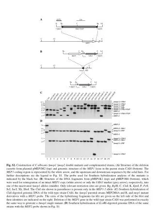
A probe 1 kb K Xh C Bg ScI 5`MEP1 URA3-FLIP 3`MEP1 C (C) C C MEP1 B Xh C Bg P ScI MEP1 TACT1 URA3 3`MEP1 C (C) C Dmep1::FRT Xh Bg P ScI 5`MEP1 TACT1 URA3 3`MEP1 C MEP12MK1A MEP12MK2A MEP12MK2B MEP12MK1B MEP12M2A MEP12M2B MEP12M5B MEP12M3A MEP12M6B MEP12M1B MEP12M3B MEP12M5A MEP12M6A MEP12M1A MEP12M4A MEP12M4B MEP2M4B MEP2M4A CAI4 10.3 Dmep1-2::URA3 8.8 Dmep1-1::URA3 8.6 Dmep1-2::FRT 7.1 Dmep1-1::FRT 6.6 Dmep1-2::URA3-FLIP 6.0 MEP1-2 5.1 Dmep1-1::URA3-FLIP 4.5 MEP1-1 D MEP2-2 5.6 MEP2-2::URA3 4.6 Dmep2-2::FRT 4.3 4.1 MEP2-1 3.1 Dmep2-2::URA3, MEP2-1::URA3 2.8 Dmep2-1::FRT 1.6 Dmep2-1::URA3 1 2 3 4 5 6 7 8 9 10 11 12 13 14 15 16 17 18 19 Fig. S2. Construction of C.albicans Dmep1 Dmep2 double mutants and complemented strains. (A) Structure of the deletion cassette from plasmid pMEP1M2 (top) and genomic structure of the MEP1 locus in the parent strain CAI4 (bottom). The MEP1 coding region is represented by the white arrow, and the upstream and downstream sequences by the solid lines. For further descriptions see the legend to Fig. S1. The probe used for Southern hybridization analysis of the mutants is indicated by the black bar. (B) Structure of the DNA fragments from pMEP1K1 (top) and pMEP1M4 (bottom), which were used for reintegration of an intact MEP1 copy (white arrow) or only the URA3 marker (grey arrow), respectively, into one of the inactivated Dmep1 alleles (middle). Only relevant restriction sites are given. Bg, BglII; C, ClaI; K, KpnI; P, PstI; ScI, SacI; Xh, XhoI. The ClaI site shown in parentheses is present only in the MEP1-1 allele. (C) Southern hybridization of ClaI-digested genomic DNA of the wild-type strain CAI4, the Dmep2 parental strains MEP2M4A and B, and mep1 mutant derivatives with a MEP1 probe. The sizes of the hybridizing fragments (in kb) are given on the left side of the blot and their identities are indicated on the right. Deletion of the MEP1 gene in the wild-type strain CAI4 was performed in exactly the same way to generate a Dmep1 single mutant. (D) Southern hybridization of EcoRI-digested genomic DNA of the same strains with the MEP2 probe shown in Fig. S1.