Download
1 / 93
930 likes | 1.01k Vues
Arboviruses are transmitted by blood-sucking arthropods, with over 500 types categorized by antigenic relationships. This article explores the taxonomy, structure, and pathogenesis of Alphavirus, a medically important arbovirus group.

E N D
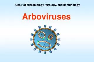
Chair of Microbiology, Virology, and Immunology Arboviruses
The arthropod-borne viruses, or arboviruses, are a group of infectious agents that are transmitted by blood-sucking arthropods from one vertebrate host to another. They can multiply in the tissues of the arthropod without evidence of disease or damage. The vector acquires a lifelong infection through the ingestion of blood from a viremic vertebrate. All arboviruses have an RNA genome, and most have a lipid-containing envelope and consequently are inactivated by ether or sodium deoxycholate. There are more than 500 arboviruses, grouped according to their antigenic relationships.
Diseases produced by the arboviruses may be divided into 3 clinical syndromes: • fevers of an undifferentiated type with or without a maculopapular rash and usually benign; • encephalitis, often with a high case fatality rate; • hemorrhagic fevers, also frequently severe and fatal. • These categories are somewhat arbitrary, and some arboviruses may be associated with more than one syndrome, eg, dengue.
Togaviridae • Genera: • Alphavirus (typical virus – Sindbis virus) • Rubivirus • Pestivirus
Togaviruses general properties Small viruses, (+) ssRNA, linear, м.w. 4,0 МD, Icosahedral symmetry, envelope, virus-specific polypeptides, some of them – glycoproteins. unstable at room temperature, stable at —70 °C, and rapidly inactivated by ether or by 1:1000 sodium deoxycholate. This property separates them easily from enteroviruses
Alphavirus There are 30 alphaviruses, 13 of them can infect human
Virus structure Virions are spherical, 60-70 nm in diameter, icosahedral nucleocapsid enclosed in a lipid-protein envelope. Alphavirus RNA is a single 42S strand of approximately 4x10⁶ daltons Virion RNA is positive sense: it can function intracellularly as mRNA, and the RNA alone has been shown experimentally to be infectious. The single capsid protein (C protein) Two envelope viral glycoproteins (E1 and E2) of molecular weights of 48,000 to 52,000 daltons. A small third protein (E3) of molecular weight 10,000 to 12,000 daltons remains virion-associated in Semliki Forest virus but is dispatched as a soluble protein in most other alphaviruses. On the virion surface, E1 and E2 are closely paired, and together form trimers that appear as "spikes" in an orderly array.
The viruses infect many cell lines, embryonated eggs, mice, birds, bats, mules, horses, and other animals. In susceptible vertebrate hosts, primary virus multiplication occurs either in myeloid and lymphoid cells or in vascular endothelium. Multiplication in the central nervous system depends on the ability of the virus to pass the blood-brain barrier and to infect nerve cells. In natural infection of birds and mammals (and in experimental parenteral injection of the virus into animals), an inapparent infection develops in a majority. However, for several days there is viremia, and arthropod vectors acquire the virus by sucking blood during this period—the first step in its dissemination to other hosts.
Classification and Antigenic Types • Classification is based upon antigenic relationships. • Viruses have been grouped into seven antigenic complexes. Typical species in four medically important antigenic complexes are: • Venezuelan equine encephalitis, • Eastern equine encephalitis, • Western equine encephalitis, • Semliki Forest viruses.
Multiplication. Alphaviruses attach to cells via interactions between E2 and cellular receptors. Entry is thought to take place in mildly acidic endosomal vacuoles where glycoprotein spikes undergo conformational rearrangements and an acid-dependent fusion event (principally a function of E1) delivers genomic RNA to the cell cytoplasm. Viral replication occurs in the cytoplasm. Initial translation of virion RNA produces a polyprotein that is proteolytically cleaved into an RNA polymerase.
Transcription of the virion RNA through a negative-strand RNA intermediate produces a 26S positive-strand mRNA (short) which encodes only the structural proteins, as well as additional 42S RNA (long), which is incorporated into progeny virions. Translation from the 26S mRNA (which represents the 3' one-third of genomic RNA) produces a polyprotein that is cleaved proteolytically into three proteins: C, PE2, and E1; PE2 is subsequently cleaved into E2 and E3. Envelope proteins formed by posttranslational cleavage are glycosylated and translocated to the plasma membrane. Virion formation occurs by budding of preformed icosahedral nucleocapsids through regionsof the plasma membrane containing E1 and E2 glycoproteins
Pathogenesis and Clinical Manifestations.Human illness caused by alphaviruses is exemplified by agents that produce three markedly different disease patterns. • Chikungunya virus is the prototype for those causing an acute (3- to 7-day) febrile illness with malaise, rash, severe arthralgias, and sometimes arthritis. • O'nyong'nyong, Mayaro, and Ross River viruses, which are closely related (antigenically) to chikungunya virus, cause similar or identical clinical manifestations; • Sindbis viruses cause similar but milder diseases known as Ockelbo (in Sweden), Pogosta (Finland), or Karelian fever (Russia).
Epidemiology. Diseases are maintained in natural ecologic cycles involving birds and, principally, bird-feeding mosquitoes such as Culex, Aedes. Eastern equine encephalitis (EEE) virus is enzootic in fresh water swamps in the eastern United States; it causes sporadic equine and rare human cases. Small human outbreaks may occur. Alphavirus transmission Virus abbreviations: Chik, chickungunya; RR, Ross River; May, Mayaro; ONN, O'nyong-nyong; SIN, Sindbis; EEE, eastern equine encephalitis; VEE, Venezuelan equine encephalitis.
Host Defenses • Differences in susceptibility between individuals and species are not easily ascribed to specific immune responses, and a variety of non-specific defense mechanisms may be important. • Alphaviruses are efficient inducers of interferon, the production of which probably plays a role in modulating or resolving infections. • Antibodies are important in disease recovery and resistance. The appearance of neutralizing antibodies in serum coincides with viral clearance, and immune serum can diminish or prevent alphavirus infection. • Although their precise roles are not clearly established, T-cell responses are also demonstrable and may contribute substantially to immunity.
Diagnosis • Blood, cerebrospinal fluid examination, section material, blood serum to include viral cultures, is critical in differentiating bacterial from viral infections, and infectious from noninfectious etiologies. • Laboratory diagnosis can be established by isolating virus from the blood during the viremic phase or by antibody determination. • Express-methods: IRHAT, IFT, ELISA • Virological diagnosis: inoculation of laboratory animals (into the brain) – suckling mice, cell cultures, embryotaned eggs. Identificarion: plaques formation, CPE, HIT, CFT, NT • Serology: paired sera (NT, CFT, HIT, IHAT, ELISA)
Control • Control of alphavirus diseases is based on surveillance of disease and virologic activity in natural hosts and, when necessary, on control measures directed at reducing populations of vector mosquitoes. These measures include: • control of larvae and adult mosquitoes, sometimes by using ultra-low-volume aerial spray techniques; in some areas, insecticide resistance (for example, resistant C tarsalis) is a major limitation to effective control. • Inactivated vaccines are used to protect laboratory workers from eastern, western, and Venezuelan equine encephalitis viruses. An effective live attenuated Venezuelan equine encephalitis vaccine has been employed extensively in equines as an epidemic control measure, and a similar vaccine is used to protect laboratory workers. • A live attenuated chikungunya vaccine has proven safe and immunogenic in investigational human trials.
Rubellaviruses Rubella (German measles) is a common mild disease characterized by a rash. It affects children and adolescents worldwide and can also affect young adults. When rubella virus infects susceptible women early in pregnancy, it may be transmitted to the fetus and may cause birth defects. Therefore, accurate diagnosis is critical in pregnancy.
Structure Rubella virus is a spherical 40- to 80-nm, positive-sense, single-stranded RNA virus consisting of an electron-dense 30- to 35-nm core surrounded by a lipoprotein envelope. The RNA has a molecular weight of about 3 X 10⁻⁶. It has 2 structural proteins: major and minor. The virus particles are generally spherical with spiky hemagglutinin-containing surface projections. It has haemolytic and neuraminidaze activities too.
Classification and Antigenic Types Rubella virus is the single member of the genus Rubivirus in the family Togaviridae. It is serologically distinct from other members of the Togaviridae, and, unlike most other togaviruses, is not known to be transmitted by an arthropod. Only one genetically stable serotype of rubella virus has been identified. Cultivation The virus reproducts in primary cell cultures of human embryo and continious cell lines. It produses CPE as other Togaviruses. Reproduction (slowly!) is in cytoplasm. Eosinophilic inclusions are formed. We can detect the viruuses in the cells by interferention test – additional infection of cell culture with ЕСНО-11 viruses. CPE
Epidemiology • Humans are the only known reservoir of rubella virus. Incubation period - 16-18 days. Mechanism of transmission: • postnatal person-to-person transmission occurring via direct or droplet contact with the respiratory secretions of infected persons, • contact (formites), • transplacental Although the early events surrounding infection are incompletely characterized, the virus almost certainly multiplies in cells of the respiratory tract, extends to local lymph nodes, and then undergoes viremic spread to target organs.
Clinical Manifestations Postnatal rubella is often asymptomatic but may result in a generally mild, self-limited illness characterized by rash, lymphadenopathy, and low-grade fever. As is the case for many viral diseases, adults often experience more severe symptoms than do children. In addition, adolescents and adults may experience a typical mild prodrome that is not seen in infected children; this occurs 1 to 5 days before the rash and characterized by headache, malaise, and fever.
Catarata Rash Glaucoma
Host Defenses Postnatal infection rapidly induces a specific immune response which provides lifelong protection against the natural disease. Neutralizing and hemagglutination-inhibiting antibodies appear shortly after the onset of rash and reach maximum levels in 1 to 4 weeks. Specific antibodies persist after infection. Cell-mediated immunity also develops in convalescence and can be detected for years following infection.
Diagnosis • Serologic studies: • detection of IgM and/or fourfold antibody rises); • presence of specific IgM antibodies indicates recent rubella infection; • Specific IgG antibodies in healthy individuals demonstrate immunity to rubella; • Antibodies are detectible by a variety of methods including: • neutralization test (seldom used because of its complexity and expense), • hemagglutination inhibition, • ELISA, • indirect immunofluorescent immunoassay. • Virologic studies: • Virus can be readily recovered in cell cultures from respiratory tract secretions and, in infants with congenital infection, from urine, cerebrospinal fluid, and blood. Presence of virus in inoculated cultures can be recognized by viral interference or immunoperoxidase staining assays. Because virus isolation procedures are costly and require a relatively sophisticated virologic laboratory, they are seldom used except for the diagnosis of congenital rubella.
Prophylaxis Live attenuated vaccines are used for vaccination of girls 12-15 months of old, revaccination –in 12-14 years of old. • ЕRЕVAX (rubella), Belgium • RUDIVAX (rubella), France • PRIORIX (rubella,measles,mumps), Belgium • TRIMOVAX (rubella, measles, mumps),France • MMR (rubella,measles, mumps), USA Immunoglobulin has been used in attempts to prevent rubella in pregnant women exposed to the virus.
Flaviviruses • Classification and Antigenic Types • Classification within the genus is based upon antigenic relationships and genetic relationships are becoming increasingly important in classification. Viruses have been grouped into several antigenic complexes typified, for example, by dissimilar viruses: • dengue, • tick-borne encephalitis, • St. Louis encephalitis, • yellow fever viruses
Flaviviruses Flavivirus virions are spherical, 50 nm in diameter, and consist of a nucleoprotein capsid enclosed in a lipid envelope. The RNA is a single 40S positive-sense strand. The virion has a single capsid protein (C) that is approximately 13,000 daltons. The envelope consists of a lipid bilayer, a single envelope protein (E) of 51,000-59,000 daltons, and a small nonglycosylated protein (M) of approximately 8,500 daltons. Only E, which is glycosylated in most flaviviruses, is clearly demonstrable on the virion surface.
Antigenic properties In nucleocapsid there is one group-specific protein. Envelope glycoprotein has haemagglutinating properties. Surface proteins provide type-specific properties. Flavivirus Tick borne encephalitis virus
Multiplication.The mechanism by which flaviviruses enter cells probably involves an interaction between the E protein and cellular receptors, followed by a post-attachment fusion event that occurs in acidic intracytoplasmic vacuoles. Naked genomic RNA is infectious if introduced into the cytoplasm. The genomic RNA is capped but not polyadenylated; it serves as mRNA for all proteins. Structural proteins are encoded at the 5' end of the genome, and nonstructural proteins (e.g., NS-1 and RNA-dependent RNA polymerase) are encoded in the 3' two-thirds. Complementary (negative-sense) RNA, made from genomic RNA, serves as a template to generate genomic RNA. Replication occurs in the cytoplasm.
Virions are formed in perinuclear regions of the cytoplasm in association with Golgi or smooth membranes. Virions appear within cytoplasmic vacuoles and appear to exit the cell as vacuoles fuse with the plasma membrane. Unlike alphaviruses, no evidence of budding has been seen in flavivirus-infected cells, and the mechanisms of virion assembly and release remain obscure. Morphogenesis of flaviviruses
Pathogenesis and Clinical Manifestations • Flaviviruses vary widely in their pathogenic potential and mechanisms for producing human disease. It is useful to consider them in three major categories: those associated primarily with: • the encephalitis syndrome (prototype: St. Louis encephalitis), • fever-arthralgia-rash (prototype: dengue fever), • hemorrhagic fever (prototype: yellow fever).
Epidemiology.The flaviviruses constitute a highly diverse genus, and their ecology is similarly varied and complex. Only the basic concepts are considered here. The mosquito-borne encephalitis viruses (St. Louis encephalitis, Japanese encephalitis, Murray Valley encephalitis, and West Nile viruses) exist primarily as viruses of birds and are transmitted by Culex mosquitoes that feed readily on birds. Flavivirus transmission. Virus abbreviations: DEN, dengue; KFD, Kyasanur Forest Disease; OMSK, Omsk hemorrhagic fever; WN, West Nile, YF, yellow fever; SLE, St. Louis encephalitis; JE, Japanese encephalitis; POW, Powssan, RSSE, Russian spring-summer encephalitis; MVE, Murray Valley encephalitis; ROC, Rocio.
Human infection with both mosquito-borne and tick-borne flaviviruses is initiated by deposition of virus through the skin via the saliva of an infected arthropod. Pathogenesis of flaviviruses.
The tick-borne flaviviruses are maintained by tick-mammal cycles and by transovarian transmission in ticks. Humans are infected with this subgroup of flavivirus through the bite of infected ticks, and thousands of cases may occur annually in the region of the Eurasian continent between central Europe and western Siberia. A closely-related virus transmitted by Ixodes ticks, termed Powassan virus, produces sporadic cases of encephalitis in the eastern parts of Canada and the U.S.
In most human infections with St. Louis encephalitis (SLE) and Japanese encephalitis (JE) viruses, there is either no apparent disease or a nonspecific febrile illness with headache. Clinical manifestations of encephalitis due to SLE, JE, or Murray Valley encephalitis (MVE) virus begin as fever, headache, and stiff neck, and progress to an altered level of consciousness and focal neurologic deficits (e.g., tremors, pathologic reflexes, cranial nerve palsies, nystagmus, ataxia). Paralysis is more commonly seen with JE and MVE.
Structure of the Yellow Fever Virus • Spherical viruses • Capsid, Membrane protein, and Envelope protein • Capsid (C protein) • Structural Protein • Houses the RNA and the viral RNA polymerase • Membrane protein (prM/M) • Helps the capsid pass through the cell membrane. • Nonstructural proteins (NS) • Nonstructural protein 1 (NS1) is a glycosylated protein found on an infected cell’s surface and is involved in viral assembly and release. This protein induces protective antibodies. • Nonstructural protein 5 is a viral RNA polymerase.
History • Carlos Finlay • Cuban physician and scientist. • Pioneered the early research on YF. • Hypothesized that YF was spread by mosquitoes. • Walter Reed • Appointed by the government to head the Yellow Fever Commission.
Geography Out of Africa Mosquito larvae carried in ship’s water barrels spread the virus to the Americas: A new cycle in South American monkeys
Transmission of Yellow Fever • Three types of transmission to humans: • Sylatic or jungle YF • Intermediate YF • Urban YF • Vertical and Horizontal Transmission • Vertical transmission is mosquito to mosquito. • Horizontal transmission is when an infected mosquito bites a non-infected human or monkey. Mosquitoes (Aedes spp. in Africa, and Haemagogus spp. in South America)
There are 2 kinds of yellow fever • One is jungle fever that is spread from monkeys to people by mosquitoes. • The other kind is found in urban areas and are spread from person to person by mosquitoes