Download
1 / 73
750 likes | 933 Vues
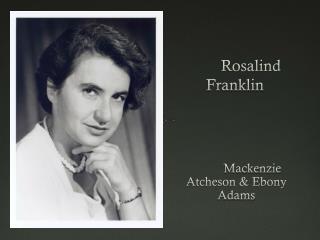
Roslyn Franklin. http://www.sdsc.edu/ScienceWomen/franklin.html. How would YOU go about determining the mechanism of DNA replication?????. What would a geneticist do?. What would a biochemist do?. Figure 5-31 Action of DNA polymerases. Page 99.

E N D
Roslyn Franklin • http://www.sdsc.edu/ScienceWomen/franklin.html
How would YOU go about determining the mechanism of DNA replication????? What would a geneticist do? What would a biochemist do?
Here’s a computer modelhttp://www.youtube.com/watch?v=4jtmOZaIvS0 Overview of DNA and replication http://207.207.4.198/pub/flash/24/menu.swf Another one with review questions http://www.wiley.com/college/pratt/0471393878/student/animations/dna_replication/index.html This is a pretty good outline: http://www.youtube.com/watch?v=teV62zrm2P0&NR=1
Paper on DNA Repairhttp://www.sciencemag.org/content/332/6036/1443.full?sid=5f09e4ee-ff8b-4f8b-acb2-e79dd986e8f6 Science 17 June 2011: Vol. 332 no. 6036 pp. 1443-1446 SIRT6 Promotes DNA Repair Under Stress by Activating PARP1 Zhiyong Mao, Christopher Hine, Xiao Tian, Michael Van Meter, Matthew Au, Amita Vaidya, Andrei Seluanov*, Vera Gorbunova*
Diseases often result from failure of DNA repair systems • colon cancer • cellular ultraviolet sensitivity • Werner syndrome (premature aging, retarded growth) • Bloom syndrome (sunlight hypersensitivity)
DNA Repair and Mutations Chemical reactions and some physical processes constantly damage genomic DNA At the molecular level, damage usually involves changes in the structure of one of the strands Vast majority are corrected by repair systems using the other strand as a template Some base changes escape repair and the incorrect base serves as a template in replication The daughter DNA carries a changed sequence in both strands; the DNA has been mutated Accumulation of mutations in eukaryotic cells is strongly correlated with cancer; most carcinogens are also mutagens
Molecular Mechanisms of Oxidative and Chemical Mutagenesis • Oxidative damage • Hydroxylation of guanine • Mitochondrial DNA is most susceptible • Chemical alkylation • Methylation of guanine • Cells have mechanisms to correct most of these modifications
Endogenous and exogenous alkylating agents (tobacco smoke, some anticancer drugs). Alkylation destabilizes the glycoside bond and can ultimately lead to backbone breaks. O6-alkylguanine has a different pattern of H-bond donor and acceptor atoms than the parent guanine base. As a result, it base pairs with T instead of C, giving rise to G A transition after the second round of replication:
Molecular Mechanisms of Spontaneous Mutagenesis • Deamination • Very slow reactions • Large number of residues • The net effect is significant: 100 C ????? • U events /day in a mammalian cell • Depurination • N-glycosidic bond is hydrolyzed • Significant for purines: 10,000 purines lost/day in a mammalian cell • Cells have mechanisms to correct most of these modifications.
Page 1173 Figure 30-51 Types and sites of chemical damage to which DNA is normally susceptible in vivo. Red, oxidation; blue, hydrolysis; green, methylation.
Some well-characterized nonenzymatic reactions of nucleotides. The resulting lesion, in which the deoxyribose is present but the base is not, is called an abasic site or an AP site (apurinic site or, rarely, apyrimidinic site). The deoxyribose remaining after depurination is readily converted from the β-furanose to the aldehyde form!!!
Molecular Mechanisms of Radiation-Induced Mutagenesis • UV light induces dimerization of pyrimidines, this may be the main mechanism for skin cancers • Ionizing radiation (X-rays and -rays) causes ring opening and strand breaking. These are difficult to fix • Cells can repair some of these modifications, but others cause mutations. Accumulation of mutations is linked to aging and carcinogenesis
DNA repair enzymes • a lot of DNA damage → elevated levels of repair enzymes • extreme change in cell's environment (heat, UV, radiation) activates genes that code DNA repair enzymes • E.g., heat-shock proteins are produced in heat-shock response to high temperatures.
Damage of the double helix • Single strand damage • information is still backed up in the other strand • Double strand damage • no backup • can cause the chromosome to break up
Types of DNA Repair Systems • Single strand repair • Nucleotide Excision Repair • Base Excision Repair • Mismatch Repair (shortly after replication) • Double strand repair • Homologous end-joining • Non-homologous end-joining
Types of DNA Repair Systems Mismatches arise from occasional incorporation of incorrect nucleotides Abnormal bases arise from spontaneous deamination reactions or via chemical alkylation (alk genes) Pyrimidine dimers form when DNA is exposed to UV light Backbone lesions occur from exposure to ionizing radiation
Formation of pyrimidine dimers induced by UV light. (b) Formation of a cyclobutane pyrimidine dimer introduces a bend or kink into the DNA
Figure 30-52 The cyclobutylthymine dimer that forms on UV irradiation of two adjacent thymine residues on a DNA strand. Photolyase: repairs cyclobutane pyrimidine dimers. Uses the energy of light to catalyze the reversal of the cyclobutane bonds, producing intact DNA. Not very important in mammals.
A large multienzyme compound scans the DNA strand for anomalies. Nucleotide-excision repair in E. coli and humans Upon detection a nuclease cuts the strand on both sides of the damage. • DNA helicase removes the oligonucleotide. • The gap is repaired by DNA polymerase and DNA ligase enzymes
Figure 30-56 Base Excision RepairDNA glycosylases hydrolyze the glycosidic bond of their corresponding altered base to yield an AP site. Page 1177
Base-excision repair pathway 1 DNA glycosylase recognizes damaged base, cleaves N-glycoside bond 2 AP endonuclease cleaves backbone near the AP site. 3 DNA pol I initiates repair synthesis from the free 3′ OH at the nick, removing and replacing the damaged strand. 4 Nick sealed by DNA ligase.
O6-alkylguanine DNA alkyltransferase (AGT) Directly repairs alkylation damage (O6-alkylguanines) by transferring the O6-alkyl group from damaged guanine in DNA to a Cys residue in the AGT active site in a stoichiometric reaction. The protein is inactivated via alkylation and undergoes proteolytic degradation. AGT protein is highly conserved: helix-turn-helix DNA binding motif the alkylated base is “flipped” out of the helix to enter the hydrophobic alkyl-binding pocket of the protein high metabolic cost for the cell is outweighed by the need to maintain genetic integrity
Binding of Proteins to DNA Often Involves Hydrogen Bonding Types of domains that bind DNA: Helix-turn-helix Zinc Finger Leucine Zipper Helix-loop-helix
Functional groups on all four base pairs that are displayed in the major and minor grooves of DNA
A = H bond acceptor D = H bond donor
Guanine-Arginine: One of the most common DNA-protein interactions. Because of its specific geometry of H-bond acceptors, guanine can be unambiguously recognized by the side chain of arginine
Helix-Turn-Helix Motif is Common in DNA-Binding Proteins One of the helixes (red) fits into the major groove of DNA Four DNA-binding helix-turn-helix motifs (gray) in the Lac repressor
Important Points: a handshake leads to a bear-hug Specific recognition of DNA targets by the helix-turn-helix motif involves interactions between sides of the recognition helix and bases in the major groove of the DNA But, specific recognition of DNA sequences is to a large extent governed by other interactions within complementary surfaces between the protein and the DNA These interactions frequently involve H-bonds from protein main-chain atoms to the DNA backbone in both the major and the minor groove and are dependent on the sequence-specific deformability of the target DNA
Helix-turn-helix. (a) DNA-binding domain of the Lac repressor The helix-turn-helix motif is shown in red and orange; the DNA recognition helix is red.
Helix-turn-helix. (c) Surface rendering of the DNA-binding domain of the Lac repressor bound to DNA.
The DNA-binding domain separated from the DNA, with the binding interaction surfaces shown. groups on the protein and DNA that interact through H-bonding groups that interact through hydrophobic interactions
Extrahelical Damaged Base Recognition by DNA Glycosylase Enzymes • Intermediates on the base flipping pathways of hOGG1 (human OxoGuanine Glycosidase) • The exo-site complex of hOGG1 with an extrahelical guanine obtained by disulfide crosslinking technology (left). The fully extrahelical complex with 8-oxoG is shown on the right for comparison. Chemistry – A European JournalVolume 14, Issue 3, pages 786-793, 15 NOV 2007 DOI: 10.1002/chem.200701501http://onlinelibrary.wiley.com/doi/10.1002/chem.200701501/full#fig5
Methylation and mismatch repair Really only understood well in E.coli. The methylation occurs at the N6 of adenines in (5′)GATC sequences. (palindrome) Dam=DNA adenine methylation
Double strand repair • Homologous end-joining • damaged site is copied from the other chromosome by special recombination proteins
Double strand repair • Nonhomologous end-joining • only in emergency situations • two broken ends of DNA are joined together • a couple of nucleotides are cut from both of the strands • ligase joins the strands together
Cell Cycle and DNA repair • Cell cycle is delayed if there is a lot of DNA damage. • Repairing DNA as well as signals sent by damaged DNA delays progression of cell cycle. This ensures that DNA damages are repaired before the cell divides
Paper on DNA Repairhttp://www.sciencemag.org/content/332/6036/1443.full?sid=5f09e4ee-ff8b-4f8b-acb2-e79dd986e8f6 Science 17 June 2011: Vol. 332 no. 6036 pp. 1443-1446 SIRT6 Promotes DNA Repair Under Stress by Activating PARP1 Zhiyong Mao, Christopher Hine, Xiao Tian, Michael Van Meter, Matthew Au, Amita Vaidya, Andrei Seluanov*, Vera Gorbunova*
Abstract Sirtuin 6 (SIRT6) is a mammalian homolog of the yeast Sir2 deacetylase. Mice deficient for SIRT6 exhibit genome instability. Here, we show that in mammalian cells subjected to oxidative stress, SIRT6 is recruited to the sites of DNA double-strand breaks (DSBs) and stimulates DSB repair, through both nonhomologous end joining and homologous recombination. Our results indicate that SIRT6 physically associates with poly[adenosine diphosphate (ADP)–ribose] polymerase 1 (PARP1) and mono-ADP-ribosylates PARP1 on lysine residue 521, thereby stimulating PARP1 poly-ADP-ribosylase activity and enhancing DSB repair under oxidative stress.
It is interesting to note that NAD+ is required as substrate for generating ADP-ribose monomers. The overactivation of PARP may deplete the stores of cellular NAD+ and induce a progressive ATP depletion, since glucose oxidation is inhibited, and necrotic cell death. In this regard, PARP is inactivated by caspase-3 cleavage (in a specific domain of the enzyme) during programmed cell death. PARP enzymes are essential in a number of cellular functions, including expression of inflammatory genes: PARP1 is required for the induction of ICAM-1 gene expression by smooth muscle cells, in response to TNF. Activity PAR is synthesized using nicotinamide (NAM) as the leaving group. This leaves a pyrophosphate as the linking group between ribose sugars rather than single phosphate groups. This creates some special bulk to a PAR bridge, which may have an additional role in cell signaling. Role in repairing DNA nicks One important function of PARP is assisting in the repair of single-strand DNA nicks. It binds sites with single-strand breaks through its N-terminal zinc fingers and will recruit XRCC1, DNA ligase III, DNA polymerase beta, and a kinase to the nick. This is called base excision repair (BER). PARP-1 is also known for its role in transcription through remodeling of chromatin by PARylating histones and relaxing chromatin structure, thus allowing transcription complex to access genes.