Download
1 / 56
600 likes | 917 Vues
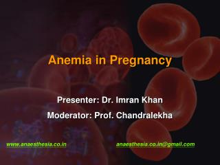
Acquired Anemia During Pregnancy. Reihaneh Pirjani , M.D. Perinatologist , Assistant Professor, Tehran University of Medical Sciences. Hematologic changes in pregnancy. Physiologic anemia Neutrophilia Mild thrombocytopenia Increased procoagulant factors Diminished fibrinolysis

E N D
Acquired Anemia During Pregnancy ReihanehPirjani, M.D. Perinatologist, Assistant Professor, Tehran University of Medical Sciences
Hematologic changes in pregnancy • Physiologic anemia • Neutrophilia • Mild thrombocytopenia • Increased procoagulant factors • Diminished fibrinolysis • Plasma volume increases • Red blood cell mass increase
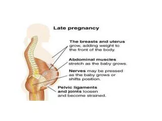
A greater expansion of plasma volume relative to the increase in hemoglobin mass and erythrocyte volume is responsible for the modest fall in hemoglobin levels.
Physiologic anemia of pregnancy should resolve by six weeks postpartum since plasma volume has returned to normal by that time.
Causes of Acquired Anemia during Pregnancy Iron-deficiency anemia(most common) Anemia caused by acute blood loss Anemia of inflammation or malignancy Megaloblastic anemia Acquired hemolytic anemia
75%از کم خونی های دوران بارداری ناشی ازفقر آهن است The two most common causes of anemia during pregnancy and the puerperium are iron deficiency and acute blood loss
If The dietary iron intake is poor The interval between pregnancies is short The delivery is complicated by hemorrhage Iron deficiency anemia readily and rapidly develops.
Anemia has defined as hemoglobin levels of : less than 11 g/dL (hematocrit less than 33 percent) in the first and third trimesters less than 10.5 g/dL (hematocrit less than 32 percent) in the second trimester
اولین قدم در بررسی آنمی در حاملگی این است که بتوانیم آنمی فیزیولوژیک را از موارد پاتولوژیک افتراق دهیم. • برای این منظور: • اولأ باید نیاز طبیعی به آهن را در طی حاملگی بدانیم. • ثانیأ باید بتوانیم ازپارامترهای آزمایشگاهی استفاده مناسب کرده یا به عبارت دیگر یافته های ازمایشگاهی را بتوانیم درست تفسیر کنیم.
In a typical singleton gestation, the maternal need for iron averages close to 1000 mg. Of this, 300 mg is for the fetus and placenta; 500 mg for maternal hemoglobin mass expansion; and 200 mg that is shed normally through the gut, urine, and skin.
patient should undergo a standard evaluation : • complete blood count • review of peripheral smear • reticulocyte count • serum Fe/TIBC, and ferritin
Classical morphological evidence of iron-deficiency anemia—erythrocyte hypochromia and microcytosis—is less prominent in the pregnant.
Classical morphological evidence of iron-deficiency anemia—erythrocyte hypochromia and microcytosis—is less prominent in the pregnant Moderate iron-deficiency anemia during pregnancy usually is not accompanied by obvious morphological changes in erythrocytes
If iron deficiency is combined with folate or vitamin BI2 deficiency, normocytic and normochromic RBCs are observed on the peripheral blood smear.
pathologic change in iron deficiency anemia The first pathologic change to occur in iron deficiency anemia is the depletion of bone marrow, liver, and spleen iron stores. The serum iron level falls. The total iron-binding capacity rises, because this is a reflection of unbound transferrin. A falling hemoglobin and hematocrit follow.
In adult women, iron stores are located in the bone marrow, liver and spleen in the form of ferritin. Ferritin constitutes approximately 25 percent (500mg) of the 2-g of iron stores found in the normal woman. Approximately 65 percent of stored iron is located in the circulating RBCs.
Ferritin • The ferritin level indicates the total status of her iron stores. • Serum ferritin levels normally decrease minimally during pregnancy.
Ferritin • Ferritin levels are variable and can change 25 percent from one day to the next. • However, a significantly reduced ferritin concentration is indicative of iron deficiency anemia and is the best parameter to judge the degree of iron deficiency.
Ferritin Levels less than 10 to 15 mg/L confirm iron-deficiency anemia
Serum Transferrin Receptor measuring serum transferrin receptors can give a better index of true iron status. A reduction in the iron supply increases TfRsynthesis. In patients with iron deficiency anemia, the plasma receptor is elevated threefold.
Serum Transferrin Receptor Ferritin can be elevated in acute and chronic infections, whereas transferrin receptors do not change in response to an infection. Also receptor concentrations are not confounded by the hemodynamic changes of pregnancy.
Serum Transferrin Receptor This test is not yet readily available but may help us in the future to detect irondeficient patients.
Serum Iron A serum iron concentration less than 60 mg/dl with less than 16 percent saturation of transferrin is suggestive of iron deficiency.
Serum Iron Asingle normal serum iron concentration does not rule out iron deficiency. For example, a patient may take iron for several days, and this may result in a transiently normal serum iron concentration while iron stores are still negligible.
Total Iron-Binding Capacity(TIBC) TIBC rises in association with iron deficiency and decreases in chronic inflammatory states. It is increased in pregnancy because of the increase in plasma volume.
Total Iron-Binding Capacity(TIBC) • An increase in iron-binding capacity is not reliable, because: • 15 percent of pregnant women without iron deficiency show an increase in this parameter.
Management Options In pregnancy, iron absorption from the duodenum increases.
Management Options Antacid medications, commonly used by many patients, decreases the absorption of iron. Chronic use of H2 blockers and proton pump inhibitors also diminishes iron absorption. Vitamin C, in addition to the iron, may increase the acid environment of the stomach and increase absorption.
Iron prophylaxis, however, is safe as only amounts that can be used are absorbed. With the exception of dyspepsia and constipation, side effects are few. If the iron is not needed, it will not be absorbed and will be excreted in the feces.
In iron-deficient patients, one iron tablet three times daily has been recommended. ferrous sulfate, fumarate, or gluconate—that provide approximately 200 mg daily of elemental iron.
There are equivalent increases in hemoglobin levels in women treated with either oral or parenteral iron therapy.
Parenteral iron is indicated in those who cannot or will not take oral iron therapy and are not anemic enough to require transfusion.
Transfusions of red cells or whole blood seldom are indicated unless hypovolemia from blood loss coexists or an emergency operative procedure must be performed on a severely anemic woman.
To replenish iron stores, oral therapy should be continued for 3 months after anemia has been corrected
Recombinant human erythropoietin has also been used in difficult cases. It does not cross the placenta but carries a risk of hypertension and thrombosis. Its role in pregnancy is not well established.
MegaloblasticAnemia megaloblastic anemia beginning during pregnancy almost always results from folic acid deficiency.
The earliest biochemical evidence is low plasma folic acid concentrations.
Early morphological changes usually include neutrophils that are hypersegmented and newly formed erythrocytes that are macrocytic.
As the anemia becomes more intense, peripheral nucleated erythrocytes appear and examination of the bone marrow discloses megaloblastic erythropoiesis
Treatment The treatment of pregnancy-induced megaloblastic anemia should include folic acid, a nutritious diet, and iron. As little as 1 mg of folic acid administered orally once daily
Treatment By 4 to 7 days after the beginning of treatment, the reticulocyte count is increased, and leukopenia and thrombocytopenia are corrected
Megaloblastic anemia during pregnancy caused by lack of vitamin B12, that is, cyanocobalamin, is exceedingly rare. During pregnancy, vitamin B12 levels are lower than nonpregnant values because of decreased levels of binding proteins
vitamin B12 deficiency in pregnant women is more likely encountered following partial or total gastric resection. Other causes are Crohn disease, ileal resection, and bacterial overgrowth in the small bowel.
Women who have had a total gastrectomy require 1000 g of vitamin B12 intramuscularly at monthly intervals. Those with a partial gastrectomy usually do not need such therapy, but vitamin B12 levels during pregnancy should be measured. .
Addisonian pernicious anemia, a lack of intrinsic factor is an extremely uncommon autoimmune disease Typically has its onset after age 40 years. Unless treated with vitamin B12, infertility may be a complication.
Anemia Associated with Chronic Disease • renal insufficiency, • suppuration, • inflammatory bowel disease, • systemic lupus erythematosus, • granulomatous infections, • malignant neoplasms, • rheumatoid arthritis
Bone marrow cellular morphology is not altered Serum iron concentration is decreased Ferritin levels usually are elevated.
Chronic Renal Disease: • Chronic renal insufficiency may be accompanied by anemia • usually due to erythropoietin deficiency • an element of anemia of chronic disease
Women who have acute pyelonephritis with sepsis often develop overt anemia. This is caused by acute red cell destruction from endotoxin-mediated sepsis, but with normal erythropoietin production
Treatment Adequate iron stores must be ensured. In pregnancies complicated by chronic renal insufficiency, recombinant erythropoietin is usually considered when the hematocrit approximates 20 percent. One worrisome side effect is hypertension, which is already prevalent in women with renal disease.