Download
1 / 18
441 likes | 1.87k Vues
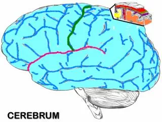
White Matter Of Cerebrum. myelinated axons that form bundles of fibers between différent nuclear masses. White Matter Of Cerebrum. Intracortical fibers short, project to nearby cortical areas most from horizontal neurons in layer I

E N D
White Matter Of Cerebrum myelinated axons thatform bundles of fibers between différent nuclear masses
White Matter Of Cerebrum • Intracortical fibers • short, project to nearby cortical areas • most from horizontal neurons in layer I • some from horizontal axon collaterals from pyramidal cells
White Matter Of Cerebrum Association fibers • gyrus to gyrus and lobe to lobe in the same hemisphere • arcuate fibers connect adjacent gyri • long association fibers connect distant gyri • originate from pyramidal neurons in layers II and III
White Matter Of Cerebrum Short Association fibres • Arcuate fibers • “U” Cortical fibres • Connect the Adjacent gyri’s
White Matter Of Cerebrum Long Association fibers Long association fibers • Connect the different lobes of the same hemisphere • Cingulum • Superior longitudinal Bundle • Inferior longitudinal Bundle • Arcuate fasciculus • Uncinate fasciculus • Superior longitudinal fasciculus: connects sup. & medial frontal gyri to parietal & occipital lobe • Arcuate fasciculus: arches around the insular region, connects Broca’s and Wernicke’s areas • Uncinate fasciculus: connects orbital frontal gyri to anterior parts of the temporal lobe
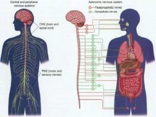
White Matter Of Cerebrum • Projection fibers connect cortex with subcortical neurons • corticofugal/efferent, project from cortex • corticopetal/afferent, project to cortex • Corticofugal project to corpus striatum, brainstem, and spinal cord • Corticopetal projections arise mainly from the thalamus - the thalamic radiations • Internal capsule carries most of these connections
Clinical Case • A patient presented with paralysis of the left side of the limbs and left side of the lower face and deviation of the tongue to the left with no atrophy and with no loss of taste sensation. This constellation of deficits most likely resulted from a lesion of the: • Left internal capsule • Right internal capsule • Left pontine tegmentum • Ventromedial medulla on the right side • Ventromedial medulla on the left side
Clinical Case • A 35-year-old man suffered a stroke that did not cause paralysis. However, he discovered that he was unable to perform complex learned movements. The region of the cerebral cortex most likely affected by the stroke was the: • Precentral gyrus • Postcentral gyrus • Premotor cortex • Temporal neocortex • Prefrontal cortex
White Matter Of Cerebrum Projection fibers are deep to association fibers • Corona Radiata: afferent & efferent fibers in radially arranged bundles that converge towards the brainstem • form a compact band of fibers (Internal Capsule)between the caudate nucleus and the thalamus • Internal Capsule: • Anterior limb: separates caudate nucleus from putamen • Posterior limb: separates thalamus from lentiform nucleus ( = putamen + globus pallidus) • Both limbs meet at the genu of the internal capsule
Internal Capsule Part 1 • Anterior limb • frontal lobe connections • cortex to striatum and pontine nuclei • anterior and medial thalamus to frontal lobe • Genu • Ventral anterior and ventral lateral thalamus to premotor and motor cortex
Internal CapsulePart 2 • Posterior limb • corticospinal • retrolenticular = optic radiations • sublenticular = auditory radia. • Somatosensory thalamocortical fibers
Internal Capsule Clinical Illustration • Posterior limb of internal capsule is most common site of stroke (supplied by MCA) • Capsular stroke • contralateral spastic hemiplegia • contralateral hemianesthesia • contralateral lower facial paralysis • if retrolenticular part is damaged then contralateral homonymous hemianopsia will result (deficit in contralateral half of visual field of both eyes) • Anterior limb supplied by ACA
White Matter Of Cerebrum Commissural fibers Corpus callosum • Connect homologous areas of the two hemispheres • Corpus Callosum • Anterior Commissure • Posterior commissure • Hebelunar commissure • Hippocampal commissure
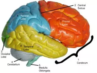
White Matter Of Cerebrum • connect homologous areas of the two hemispheres • Corpus callosum: rostrum, genu, trunk, splenium • rostrum & genu connect frontal lobes • trunk connects posterior frontal lobes, parietal lobes, and superior temporal lobe • splenium connects the occipital lobes • Originate with pyramidal neurons in layers II and III
Split Brain Patients • Transection of corpus callosum sometimes performed for epilepsy treatment • Separates 2 hemispheres except for anterior and posterior commissures
White Matter Of Cerebrum • Anterior commissure connects the inferior and middle temporal gyri in opposite hemispheres; also olfactory connections • Posterior commissure carries fibers from the pretectal nuclei and other nearby neurons