Download
1 / 12
140 likes | 601 Vues
Esophagus Dr. Nimir Dr. Safaa. Objectives. Define esophagus. Enlist relations of esophagus. Describe the constrictions of the esophagus. Give its blood and nerve supply. ESOPHAGUS. The esophagus is a muscular

E N D
Esophagus Dr. Nimir Dr. Safaa
Objectives • Define esophagus. • Enlist relations of esophagus. • Describe the constrictions of the esophagus. • Give its blood and nerve supply.
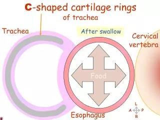
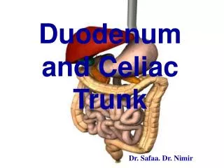
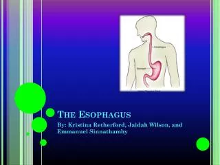
ESOPHAGUS • The esophagus is a muscular tube about 25-27cm long which connects the pharynx with the stomach (40-45 cm from teeth to cardia of the stomach). The esophagus takes a straight course through the mediastinum of the thorax and pierces the diaphragm at the esophageal hiatus to enter the abdomen and the stomach.
The Oesophagus 25-27 cm Cervical Thoracic oesophageal hiatus (T 10) Abdominal Cardia
Relations of :esophagus Posterior: vertebrae, thorathic duct, hemiazygos, accessory hemiazygos, descending aorta, first 2 intercostal arteries from aorta. Anterior: trachea to T4/T5, reccurent laryngeal nefrves, left bronchus, left atrium, diaphragm. Left: thoracic duct, aorta, left subclavian artery, lung. Right: lung, azygos vein( hence good side to approach the oesophagus surgically).
Relations of esophagus: • Posterior: vertebrae, thoracic duct, hemiazygos, accessory hemiazygos, descending aorta, first 2 intercostal arteries from aorta. • Anterior: trachea to T4/T5, reccurentlaryngeal nerves, left bronchus, left atrium, diaphragm. • Left: thoracic duct, aorta, left subclavian artery, lung. • Right: lung, azygos vein( hence good side to approach the esophagus surgically).
0 15 25 40
Blood Supply Arteries the inferior thyroid arteries the oesophageal branches of aorta left gastric artery
Veins Inferior thyroid veins Azygos and hemiazygos Left gastric vein and the inferior phrenic veins
Nerve supply left vagus --> anterior right vagus --> posterior
Clinical Notes • Esophageal Constrictions: • The esophagus has three anatomic and physiologic constrictions. The first is where the pharynx joins the upper end, the second is where the aortic arch and the left bronchus cross its anterior surface, and the third occurs where esophagus passes through diaphragm. Swallowed foreign bodies can lodge,difficultto pass an esophagoscope, strictures develop here after the drinking of caustic fluids, common sites of carcinoma of the esophagus. Remember (15 cm), (25 cm), and (40 cm). • Portal Systemic Venous Anastomosis: • At lower third of esophagus, esophageal tributaries of azygos veins (systemic veins) anastomose with esophageal tributaries of left gastric vein (which drains into the portal vein). Should the portal vein become obstructed, as in cirrhosis of the liver, portal hypertension develops, resulting in the dilatation and varicosity of the portal systemic anastomoses. Varicosed esophageal veins may rupture during the passage of food, causing hematemesis (vomiting of blood), which may be fatal. • The Esophagus and the Left Atrium of the Heart: • The relationship help to assess size of left atrium in cases of left-sided heart failure.