
Colposcopy
Colposcopy. Dr. Mounir M. Fawzy El-Hao Prof. of Ob/Gyn Ain Shams University Cairo - Egypt. What is colposcopy?. Colposcopy is visual examination of the lower genital tract (cervix, vulva, and vagina) under magnification.

Colposcopy
E N D
Presentation Transcript
Colposcopy Dr. Mounir M. Fawzy El-Hao Prof. of Ob/Gyn Ain Shams University Cairo - Egypt
What is colposcopy? • Colposcopy is visual examination of the lower genital tract (cervix, vulva, and vagina) under magnification. • It allows assessment of lesions that are difficult to be evaluated by the naked eye, especially with cervical lesions. • Colposcope is a stereoscopic binocular and the magnification power varies as 10x to 60x. Prof. Dr. Mounir M. F. El-Hao
THE COLPOSCOPE. Prof. Dr. Mounir M. F. El-Hao
The squamous epithelium: - It is continuous with the vaginal epith. - It is responsive to oestrogenic stimuli. - Normally, it is non-keratinised. -The cellular components are termed: - basal cells. - parabasal cells. - intermediate cells. - superficial cells.
* It is essential to understand the anatomy, histology of the cervix and the changes that occur with age, menstrual cycle and pregnancy. * Lack of that knowledge can cause: 1. unnecessary treatment of a normal variation of the cervix (e.g. the wrongly called cervical erosion). 2. missing a severe lesion misdiagnosed as a normal cervix.
*Oestogen lack ( absolute or relative) leads to arrest of maturation, stratification and differentiation and most cells appearing in a smear would be either intermediate ( in pregnancy) or parabasal (postmenaupausal). *This is likely to be associated with changes in the colposcopic appearances as well.
The squamous epithelium: The component cells under go a continuous process of: - Differentiatio- Maturation Stratification from the basal to the superficial cells.
Transformation zone; **The redness is due to the thinner col. epith. showing the underlying vasculature and the roughness is due to the pappillary nature. **The process of eversion occurs during the neonatal period, in pregnancy and in oral contraceptive pills users.
**Endocervical crypts can reach a depth of 10 mm from the surface, a point which should be remembered when treating CIN. **Endocervical cells appear in sheets in the cervical smear and their presence is a must for a smear to be adequate.
Squamous metaplasia: - The everted col. epith., being exposed to the acidic hostile vaginal secretions, undergoes a process of transformation to the more resilient and protective sq. epith. - It’s a process of gradual loss of col. cells and and replacement with sq. cells in 3 stages: i. Reserve cell hyperplasia. ii. Immature squamous metaplasia. iii. Mature squamous metaplasia.
SQUAMOUS METAPLASIA. Prof. Dr. Mounir M. F. El-Hao
**Metaplasia can involve surface col. epith. as well as crypts of the endocervix and can confuse pathologists with invasive cervical cancer. **Immature metaplasia can be confused with CIN for the unexperienced colposcopist.
Congenital transformation zone: - During embryogenesis, the cuboidal epith. of the canalised vaginal tube is replaced with sq. epith. sarting distally and proceeding up. - The process completes normally when the vagina and the ectocervix are covered by sq. epith. - This process can be arrested resulting cuboidal epith. at the upper vagina and on the ectocervix that will undergo sq. metaplasia later on in intrauterine life or after birth. -The area of arrested conversion and later metaplasia is called CTZ
Indications • Suspected malignancy of the cervix: • Clinically suspicious lesions. • Abnormal cervical smear. • It is also valuable in directed biopsy. • Suspected malignancy of the vagina or the vulva. • Follow-up of cases treated for pre-malignant and malignant conditions. Prof. Dr. Mounir M. F. El-Hao
Procedure • Place the patient in lithotomy position. • Clean the cervix with normal saline to remove mucoid secretions. • The lens is focused to the desired area. • Look for any pathology and for white patches. • Apply acetic acid 2-5%. The acetic acid withdraws water from cells and areas with a high nuclear density. • A green filter could be used for better visualization of the blood vessels (appear black). Prof. Dr. Mounir M. F. El-Hao
Findings • Satisfactory examination: • Should visualize the full squamo-columnar junction. • Unsatisfactory examination: • The whole lesion or the whole squamo-columnar junction can not be visualized. Prof. Dr. Mounir M. F. El-Hao
Normal Findings Prof. Dr. Mounir M. F. El-Hao
Normal squamous epithelium: • Appears smooth and pink on the portio-vaginalis. • Columnar epithelium: • Red papillary grape-like. • Transformation zone: • The junction between the columnar and squamous epithelium. • It has areas of: • Columnar epithelium. • Areas of metaplasia (immature squamous epithelium). • Glands openings. • Nabothian follicles. • Normal blood vessels branch like a tree. Prof. Dr. Mounir M. F. El-Hao
Abnormal Findings Prof. Dr. Mounir M. F. El-Hao
Atypical transformation zone: • Leukoplakia: • A white area before the application of the acetic acid. • It represents the keratin layer. • It may or may not be associated with abnormal cells. • Acetowhite epithelium: • White area after application of acetic acid. • Mosaic areas: • Polygonal white areas separated by red lines. • They are due to newly formed capillaries. • Punctuation: • Seen as rod dots stippling representing capillary loops. • Abnormal blood vessels: • Comma-like, spaghetti-like, corkscrew and hairpin vessels. Prof. Dr. Mounir M. F. El-Hao
Suspicious of invasive carcinoma: • The tissues show raised edges, irregular contour and abnormal blood vessels. Prof. Dr. Mounir M. F. El-Hao
Delineation the area of abnormality • The application of 5% acetic acid to the cervix improves visualization. • It produces a whitened area outlining the position of CIN. • This epithelium often produces a mosaic or punctate pattern, because of its vascular change. • The whole area of abnormality should be seen. • The squamo-columnar junction is the most important area to be seen. • That area often migrates up the cervical canal in postmenopausal women, and is not clearly seen. • Cone biopsy is therefore often required in postmenopausal women. Prof. Dr. Mounir M. F. El-Hao
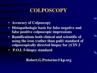
Colposcopically directed biopsy • Punch biopsy is taken from suspicious areas. • The coploscope limits the need for cone biopsy of the cervix, which has a high morbidity rate. It also limits the false negative results. • Several biopsies may be needed from the most pronounced areas. Prof. Dr. Mounir M. F. El-Hao
Thank You


![Colposcopy Market Size, Share, Trends & Growth [2018-2023]](https://cdn5.slideserve.com/9788855/colposcopy-market-worth-740-1-million-by-2023-dt.jpg)

