Download
1 / 33
340 likes | 1.21k Vues
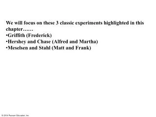
We will focus on these 3 classic experiments highlighted in this chapter…… Griffith (Frederick) Hershey and Chase (Alfred and Martha) Meselsen and Stahl (Matt and Frank). Griffith There are unknown heritable substances… Turned to a bacterial pathogen… Streptococcus pneumoniae

E N D
We will focus on these 3 classic experiments highlighted in this chapter…… • Griffith (Frederick) • Hershey and Chase (Alfred and Martha) • Meselsen and Stahl (Matt and Frank)
Griffith There are unknown heritable substances… Turned to a bacterial pathogen…Streptococcus pneumoniae Is it gram positive or negative? http://en.wikipedia.org/wiki/File:Fred_Griffith_and_%22Bobby%22_1936.jpg
Experiment Mixture of heat-killed S cells and living R cells Living S cells (control) Heat-killed S cells (control) Living R cells (control) Figure 13.2 Results Mouse dies Mouse dies Mouse healthy Mouse healthy Work by Avery identified the transforming substance as DNA Living S cells
Transformation-did not really understand mechanism • Can we do this-pick up DNA from our environment?
Hershey and Chase (1952) Their work pointed to DNA rather than proteins… Bacteriophages what are they???? (worked with one called T2)
Some phages grown in media for a couple hrs with radioactive Sulphur…(which should be incorporated into some proteins Methionine, Cysteine) Other phages grown in media for a couple hrs with radioactive Phosphorus….(which should be incorporated into DNA)
Figure 13.4 3 2 1 4 4 Experiment Batch 1: Radioactive sulfur (35S) in phage protein Centrifuged cells form a pellet. Labeled phages infect cells. Agitation frees outside phage parts from cells. Radioactivity (phage protein) found in liquid Radioactive protein Centrifuge Pellet Batch 2: Radioactive phosphorus (32P) in phage DNA Radioactive DNA Centrifuge Radioactivity (phage DNA) found in pellet Pellet
Watson-Crick Model predicted…. Each of two daughter molecules would have one parental strand and one newly made! Meselson and Stahl-clever experiment…What did they do??
Figure 13.11 3 1 2 4 Experiment Bacteria cultured in medium with 15N (heavy isotope) Bacteria transferred to medium with 14N (lighter isotope) Results Less dense DNA sample centrifuged after first replication DNA sample centrifuged after second replication More dense Conclusion First replication Second replication Predictions: Conservative model Semiconservative model Dispersive model
Watson and Crick http://www.ted.com/talks/james_watson_on_how_he_discovered_dna.html Figure 13.1
What do these terms refer to… How does this replication thing work?? • origin of replication • helicase • topoisomerase • replication fork • primase and the RNA primer • single stranded binding proteins • DNA polymerase • Search online for stronger and weaker video clips-which one is the very best and the very worst? • Email me the links to your very best and worst (with your group members names) • Jot down on the board enough of the web address that we can distinguish which ones are the same
Figure 13.7b 5 end 3 end 1 T A 2 G C G C T A 3 end 5 end
Figure 13.12 Primase Topoisomerase 3 RNA primer 5 3 5 Replication fork 3 Helicase 5 Single-strand binding proteins
Figure 13.15 Overview Leading strand Origin of replication Lagging strand Primer Leading strand Lagging strand Overall directions of replication Origin of replication 3 5 5 RNA primer 3 Sliding clamp 3 DNA pol III Parental DNA 5 3 5 Continuous elongation in the 5 to 3 direction 5 3 3 5
Figure 13.16a Overview Lagging strand Origin of replication Leading strand Lagging strand Leading strand Overall directions of replication What is going to latch on at #1?
Figure 13.16b-1 1 Primase makes RNA primer. 3 5 3 Template strand 5
Figure 13.16b-2 1 2 Primase makes RNA primer. 3 5 3 Template strand 5 DNA pol III makes Okazaki fragment 1. RNA primer for fragment 1 3 5 3 5
Figure 13.16b-3 2 1 3 Primase makes RNA primer. 3 5 3 Template strand 5 DNA pol III makes Okazaki fragment 1. RNA primer for fragment 1 3 5 3 5 DNA pol III detaches. 3 Okazaki fragment 1 5 3 5 Where is DNA pol III going to go next??
Figure 13.16c-1 4 RNA primer for fragment 2 Okazaki fragment 2 5 DNA pol III makes Okazaki fragment 2. 3 3 5 Now you have all these bits what has to happen next? And who does that?
Figure 13.16c-2 4 5 RNA primer for fragment 2 Okazaki fragment 2 5 DNA pol III makes Okazaki fragment 2. 3 3 5 5 DNA pol I replaces RNA with DNA. 3 3 5
Figure 13.16c-3 4 6 5 RNA primer for fragment 2 Okazaki fragment 2 5 DNA pol III makes Okazaki fragment 2. 3 3 5 5 DNA pol I replaces RNA with DNA. 3 3 5 DNA ligase forms bonds between DNA fragments. 5 3 3 5 Overall direction of replication
Figure 13.16 4 3 2 1 5 6 Overview Lagging strand Origin of replication Leading strand Lagging strand RNA primer for fragment 2 Leading strand Overall directions of replication Okazaki fragment 2 5 DNA pol III makes Okazaki fragment 2. 3 Primase makes RNA primer. 3 3 5 3 Template strand 5 5 5 DNA pol I replaces RNA with DNA. 3 DNA pol III makes Okazaki fragment 1. RNA primer for fragment 1 3 3 5 5 3 DNA ligase forms bonds between DNA fragments. 5 5 3 DNA pol III detaches. 3 Okazaki fragment 1 3 5 5 3 Overall direction of replication 5
Figure 13.17 Overview Origin of replication Leading strand template Lagging strand Leading strand Single-strand binding proteins Lagging strand Leading strand Overall directions of replication Leading strand Helicase DNA pol III 5 Primer 3 5 Primase 3 3 Lagging strand Parental DNA DNA pol III 5 DNA ligase DNA pol I Lagging strand template 3 5 3 5
Figure 13.17a Overview Origin of replication Leading strand Lagging strand Lagging strand Leading strand Overall directions of replication
Figure 13.17b Leading strand template Single-strand binding proteins Leading strand Helicase DNA pol III 5 Primer 3 5 Primase 3 3 Parental DNA Lagging strand template
Figure 13.17c Lagging strand DNA pol III 5 DNA pol I DNA ligase 3 5 3 5
Figure 13.14 Template strand New strand 5 5 3 3 Sugar T T A A Base Phosphate C G C G DNA poly- merase G C G C 3 T A A T P P P P i 3 P C C Pyro- phosphate Nucleotide 5 5 2 P i
Question 1. True of Leading strand, Lagging strand, or Both???? Daughter strand elongates away from replication fork Synthesizes 5’ to 3’ Multiple primers needed Made in segments Made continuously Daughter strand elongates toward replication fork
True of Leading strand, Lagging strand, or Both???? Daughter strand elongates away from replication fork Lag Synthesizes 5’ to 3’ Both Multiple primers needed Lagg Made in segments Lag Made continuously Lead Daughter strand elongates toward replication fork from Lead
Question 2. The diagram below shows a replication bubble with synthesis of the leading and lagging strands on both sides of the bubble. The parental DNA is shown in dark blue, the newly synthesized DNA is light blue, and the RNA primers associated with each strand are red. The origin of replication is indicated by the black dots on the parental strands. Rank the primers in the order they were produced. If two primers were produced at the same time, overlap them.
Question 3. The lagging strand is synthesized as a series of segments called Okazaki fragments Fragment A is the most recently synthesized and Fragment B will be synthesized next in the space between primers A and B. -----Start DNA polymerase III binds to 3’ end of primer B A. DNA polymerase I replaces primer with DNA B. DNA polymerase I binds to 5’ end of primer A C. DNA polymerase III moves 5’ to 3’ adding DNA nucleotides to primer B D. DNA ligase links fragments A and B
In an analysis of the nucleotide composition of DNA, which of the following will be found? A = G and C = T G + C = T + A A = C A + C = G + T
Cytosine makes up 42% of the nucleotides in a sample of DNA from an organism. Approximately what percentage of the nucleotides in this sample will be thymine? 31% 42% 8% 16% It cannot be determined from the information provided.