
Assessment of Mitochondrial Membrane Potential Loss in Jurkat Cells Under Different Treatments
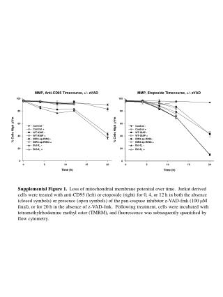
This study examines the loss of mitochondrial membrane potential over time in Jurkat-derived cells treated with anti-CD95 and etoposide. Cells were analyzed at 0, 4, or 12 hours after treatment, with and without the pan-caspase inhibitor z-VAD-fmk (100 μM final concentration), and also after a 20-hour incubation without z-VAD-fmk. Mitochondrial membrane potential was assessed using tetramethylrhodamine methyl ester (TMRM), and fluorescence quantification was performed via flow cytometry to determine treatment effects on cellular health.

Assessment of Mitochondrial Membrane Potential Loss in Jurkat Cells Under Different Treatments
E N D
Presentation Transcript
Supplemental Figure 1. Loss of mitochondrial membrane potential over time. Jurkat derived cells were treated with anti-CD95 (left) or etoposide (right) for 0, 4, or 12 h in both the absence (closed symbols) or presence (open symbols) of the pan-caspase inhibitor z-VAD-fmk (100 mM final), or for 20 h in the absence of z-VAD-fmk. Following treatment, cells were incubated with tetramethylrhodamine methyl ester (TMRM), and fluorescence was subsequently quantified by flow cytometry.