Download
1 / 23
240 likes | 710 Vues
Thromboembolic disease in pregnancy. Objectives. List the predisposing factors for thromboembolism in pregnancy Discuss the clinical presentation and management of superficial thrombophlebitis Discuss the clinical presentation and management of deep vein thrombosis

E N D
Objectives • List the predisposing factors for thromboembolism in pregnancy • Discuss the clinical presentation and management of superficial thrombophlebitis • Discuss the clinical presentation and management of deep vein thrombosis • Discuss the clinical presentation and management of pulmonary embolism in pregnancy
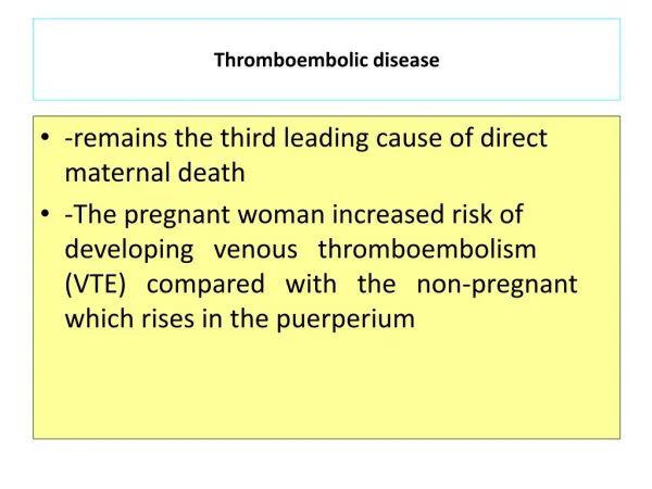
Introduction • Venous TED is one of the major causes of direct maternal deaths. Those who survive suffer significant morbidity • 2-4 fold increase compared to non-pregnant state • Cesariandelivery > vaginal delivery • 75% of DVT occur antepartum (equally distributed among all three trimesters) • 43-60% of PE occur after delivery during the first 2 weeks and in 80% of cases it is left-sided • PE is the major non-obstetric cause of maternal mortality • 2/100 000 pregnancies Fatality raten15%
Why pregnancy is associated with increased tendency for clotting ? • Venous stasis • Increased production of clotting factors V, VIII, Von Willebrand, fibrinogen • Decreased anticoagulants protein S and antithrombin • Decreased fibrinolytic activity via increased plasminogen activator inhibitor • Endothelial damage during preg and delivery
Risk factors for TED • Age over 35 yrs • Multi parity ( ≥ 4) • Obesity ( over 80 kg) • PET • Immobility • Infections • Pelvic or leg trauma • Smoking • Atrial fibrillation • Personal or family H/O TED • Thrombophilia (antithrombindefficiency, factor V Leiden, protein C, protein S DEFF.) • Antiphospholipid antibodies and lupus anticoagulant • Operative delivery (em. C/S > elective ) • Previous history of IUFD, early PET, severe IUGR, abruption
Types of venous thrombosis • Superficial thrombo phlebitis • Calf (below knee)deep vein thrombosis • Proximal or ilio-femoral deep venous thrombosis--- 70% of DVT in preg
Diagnosis • Clinical diagnosis is difficult and inaccurate in over 60% of cases of TED • Leg symptoms (oedema and pain) and dyspnea are common in pregnancy/ mimic symptoms of DVT/PE • Tachycardia may be a normal physiologic response.
Superficial thrombophlebitis • The condition is misnamed. It is not infective. the redness surrounding the affected vein is a reaction to clot • It is the commonest form of venous thrombosis in pregnancy & puerperium. It occurs in about 1% of patients and nearly always arise in existing varicose veins • The diagnosis is clinically obvious (tenderness, erythema, palpable cord-like veins)
Treatment is usually symptomatic with compression bandage, leg elevation and to encourage mobility • In some pt’s DVT need to be excluded as it may co-exist with it . Even more extension to involve deep veins rarely occurs
Calf deep venous thrombosis (CVT) • The most common clinical features are pain, local tenderness, swelling, change in skin colour and temperature • Most of CVT resolve spontaneously (75-80%) and run a benign course except when the thrombus spreads up to involve the proximal deep veins (20-25%) in which case there is 50% risk of pulmonary embolism
Proximal/ Iliofemoral DVT • It occurs more commonly than CVT and over 80% is left-sided • Symptoms are more dramatic with pain and swelling involving the entire limb • If the arterial supply is unimpaired, the leg appears swollen, blue & warm. On the other hand if arterial spasm occurs secondary to irritation from the nearby clotted vein, the leg becomes swollen, painful, white & cold
Investigations for DVT • Contrast venography • Duplex ultrasonography /commonly used with a sensitivity and specificity of 97% • Compression ultrasonography • MRI --- sensitivity and specificity 100% in nonpregnantPt • Pelvic vein ultrasound, CT scan and MRI are all tests that can be used to look for pelvic clot. • D dimer test not useful in pregnancy because it normally increases with gestational age
Pulmonary embolism (PE) • A high index of suspicion is always needed for the diagnosis of PE especially in patients with DVT or risk factors for VTE • The maternal mortality rate from untreated PE is 13% with the majority within 1 hr of the event • With early diagnosis & treatment, the survival rate is between 92-95%
The common symptoms & signs of PTE • Tachypnoea • Dyspnoea • Haemoptysis • Pleuritic chest pain • Tachycardia • Cyanosis • Pyrexia • Syncope or varying degree of shock These S &S are non-specific and in most cases there is no prior clinical evidence of DVT
Investigations for suspected PTE • Chest X- ray • ECG • Blood gases • Compression duplex Doppler to exclude DVTVentilation-perfusion isotope lung scan (V/Q) • Helical or spiral CT scan is regarded superior to V/Q scan • Spiral CT • Arteriography • CT angiography
Risk of radiologic procedures to the fetus • Radiation exposure of up to 0.05 Gy (5 rad) in utero: • Oncogenicity • Relative risks of 1.2-2.4 • Absolute risk of malignancy (baseline) in fetus is estimated to be 0.1%. • Tetratogenicity • No increase in pregnancy loss, growth or mental retardation
Treatment of acute phase TED • Standard heparin IV or the more preferred LMWH S.C should be started once the diagnosis is clinically suspected until excluded by objective testing • Treatment aims at achieving APTT 2-2.5 the control for 5-7 days then continue with prophylactic dose generally for 6-12 weeks post-nataly • For PE it should be continued for 4-6 months postnataly
Heparin is the anticoagulant of choice in pregnancy. It does not cross the placenta and in overdose action can be reversed by protamin sulphate • Osteoporosis & thrombocytopenia are complications of prolonged heparin treatment. Therefore platelet count should be monitored regularly
Legs should be elevated & graduated elastic compression stocking should be worn to reduce oedema • In DVT, calf circumference should measured daily to help monitoring the response to treatment • Massive PE requires ICU & multi disciplinary team approach • Recurrent PE may require inferior vena cava filter
Thrombolytic therapy in PE should only be given with haematologist agreement • Thoracotomy with embolectoy may be life saving • Heparin thrombo -prophylaxis has to be considered in the subsequent pregnancies or if additional risk factors appear
Oral anticoagulants • Cross the placenta and are potentially teratogenic at any stage of pregnancy • Complications of warfarin includes, nasal hypoplasia, depressed nasal bridge, irregular bone growth & intracranial fetal haemorrhage • However , they can be given after delivery and are safe for lactation
conclusion • Thrombo-embolism is amajor cause of maternal mortality &morbidity worldwide • Clinical diagnosis is unreliable but once strongly suspected, treatment should be started until objectively excluded • Dupplex Doppler, x-ray venogram & V/Q scan are the main diagnostic tools
During pregnancy, LMWH is the preferred anticoagulant as it is more effective and safer than standard heparin • Oral anticoagulants should not be given at any stage during pregnancy but they are safe & may be more convenient after delivery • High clinical suspicion with early full anticoagulation and objective diagnosis are the best ways to minimize maternal M&M and avoiding risks of the unnecessary treatment