Cochlear Implant
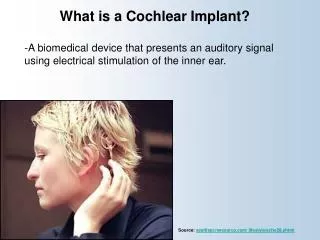
Cochlear Implant. The Fundamental Concept of Cochlear Implant. To bypass the damaged hair cells. History:. Old generation: Sound awareness only New generation: Improved communication abilities (auditory cues with lip reading, open set speech)

Cochlear Implant
E N D
Presentation Transcript
The Fundamental Concept of Cochlear Implant To bypass the damaged hair cells.
History: • Old generation: Sound awareness only • New generation: Improved communication abilities (auditory cues with lip reading, open set speech) • Since 1972 more than 16 different cochlear implants • 1984 FDA approval for adults • 1990 children approval
Anatomy Anatomy
Anatomy Scala tympani Scala vestibuli Cochlear duct Basilar membrane Vestibular membrane Tectorial membrane Hair cells (outer/inner) Cochlear nerve fibers
Sensorineural Hearing Loss Death of hair cells vs. ganglion cells Otte, et al estimated we need 10,000 ganglion cells with 3,000 apically to have good speech discrimination Apical ganglion cells tend to survive better
Structure of Cochlear Implant • External components • Internal components
Neural Responses to Sound • Temporal coding: Provide information about timing cues (rhythm and intonation. • Place coding: Rely on the tonotopic organization of a neural fibers. • Provide information about quality (timber of a speech signal – sharp to dull)
Site of Stimulation • Extracochlear • Intracochlear • Retrocochlear (lateral recess of the fourth ventricle over the cochlear nuclei.
Number of Channels • Single channel – no place coding • Multi channel
Electrode Design • 1. Single electrode • 2. Multielectrode
Indication for Cochlear Implant • Adults • 18 years old and older (no limitation by age) • Bilateral severe-to-profound sensorineural hearing loss (70 dB hearing loss or greater with little or no benefit from hearing aids for 6 months) • Psychologically suitable • No anatomic contraindications • Medically not contraindicated
Indications for Cochlear Implantation -- Children • 12 months or older • Bilateral severe-to-profound sensorineural hearing loss with PTA of 90 dB or greater in better ear • No appreciable benefit with hearing aids (parent survey when <5 yo or 30% or less on sentence recognition when >5 yo) • Must be able to tolerate wearing hearing aids and show some aided ability • Enrolled in aural/oral education program • No medical or anatomic contraindications • Motivated parents
Factors Affecting Patient Selection • Onset of deafness (congenital or adventitious) • Year of deafness • Length of sensory deprivation (i.e. no hearing aids) • Socioeconomic factors • Educational level • Individual ability to use minimal cues • General health
Factors Affecting Pt. (cont.) • Personality • Willingness to participate in rehabilitation program • Language skills • Appropriate expectations • Desire to communicate in a hearing society • Psychological stability • Cochlear patency
Audiologic Evaluation • Pure tone audiometry under headphones • Audiometry with a hearing aid in a monitored free field • Immittance testing • Speech recognition testing • OAE
Audiologic Evaluation (cont.) • Environmental sounds (closed and open set) • Speech reading (lip reading) ability • Electrical response audiometry • Auditory discrimination • Transtympanic electrical stimulation (promontory or round window test)
Medical Evaluation • Clinical history and initial interview • Preliminary examination • Complete medical and neurologic examination • Cochelar imaging using computed tomography (CT or magnetic resonance imaging (MRI) • Vestibular examination (electronystagmography) • EKG • Psychologic or psychiatric assessment or both • Vision testing • Assessment for anesthetic procedures
Contraindications • Incomplete hearing loss • Neurofibromatosis II, mental retardation, psychosis, organic brain dysfunction, unrealistic expectations • Active middle ear disease • CT findings of cochlear agenesis (Michel deformity) or narrow IAC (CN8 atresia) • Dysplasia not necessarily a contraindication, but informed consent is necessary • H/O CWD mastoidectomy • Labyrinthitisossificans—follow scans • Advanced otosclerosis
Rehabilitation 1-6-12 program Binaural Hearing Aid
Who’s eligible? • Currently: • Adults: severe to profound sensorineural hearing loss in both ears • Children (below age 2): a profound sensorineural hearing loss in both ears • Age 12 months or older • Receive little or no benefit from hearing aids • Adults: <50% open-set sentences • Children: <30% • No medical contraindications • High motivation and appropriate expectations • * Access to education and rehabilitation follow-up.
Surgical Procedure All electrode insertions are carried out through the facial recess approach. Various incision designs are used to allow wide exposure of the mastoid and squamous portions of the temporal bone. The temporalis muscle and periosteum are widely stripped to accommodate a “table” for the pedestal of the Ineraid device or the receiver-stimulator of the other devices. The mastoidectomy is not widely saucerized, but instead overhanging ledges are purposefully maintained. Care must be exercised so as not to damage the fibrous annulus during the facial recess approach..
Complications: A. Intraoperative 1. Intraoperative cannot be placed appropriately. 2. Insertion trauma 3. Gusher
Complications (cont.): B. Postoperative 1. Postauricular flap edema, necrosis or separation 2. Facial paralysis 3. Transient vertigo is more likely to occur on a totally nonfunctioning vestibular system. 4. Pain is usually associated with stimulation of Jacobson’s nerve, the tympanic branch of the glossopharyngeal nerve. 5. Facial nerve stimulation 6. Meningitis 7. Device extrusion
Rehabilitation Tuning or mapping of the external processor to meet individual auditory requirements after 3 - 4 weeks postop.