Download

1 / 7
70 likes | 183 Vues
This case report presents a unique occurrence of two distinct subtypes of meningiomas—chordoid and transitional—arising in the same region of a 64-year-old Caucasian woman. The patient presented with headaches and seizures, and imaging revealed two hyperdense lesions. Surgical intervention involved a widened pterional approach, where the chordoid meningioma was identified as WHO Grade II, while the transitional meningioma was classified as WHO Grade I. The patient remains disease-free at nine months follow-up, with no recurrence noted on imaging.

E N D
Different subtypes meningiomas arising in the same region M. Neroni, R. Gazzeri, G. Ricci e S. Esposito U.O.D. Neurosurgery, San Giovanni – Addolorata Hospital Rome
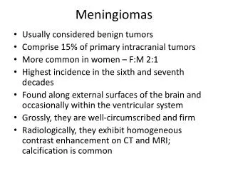
Introduction Occurrence of multiple primary brain tumors is uncommon - Chordoid meningioma is a rare isthological entity The association of two different subtypes meningiomas in the same region have not yet been reported
Case Report A 64 y.o. cauc. woman presenting with headaches since 5 months - Referred to our E.R. after acute onset of seizure No neurological deficit were disclosed
Case Report Emergency CT showed a huge pterional hyperdense lesion MR revealed two distinct lesions showing a different contrast enhancement
Widened Pterional approach Removing the anterior firm and larger tumor (intra-op histo exam revealed a chordoma) WHO II Bone and muscle were infiltrated (Simpson I) Cleavage between the lesions Posterior mass was easily removed (intra-op histo exam revealed a transitional meningioma) WHO I
Histology Anterior tumor: Chordoid Meningioma (Grade II WHO) - Posterior mass: Transitional Meningioma (Grade I WHO)
Conclusion At nine months F.U. patient is still free disease No radiotherapy was delivered - Control MR showed no recurrence