Download
1 / 26
260 likes | 395 Vues
This presentation discusses the limitations of current methods for visualizing brain variability and introduces an innovative approach using superquadric tensor glyphs for more interactive and informative visualizations. By analyzing MRI brain scans of 40 subjects, the team achieved a detailed mapping of anatomical variability, necessary for understanding functional differences across individuals. This method enhances the representation of brain structure alterations in diseases and aids in refining hypotheses about variability. Comparative evaluations show improved discrimination over traditional ellipsoid glyphs.

E N D
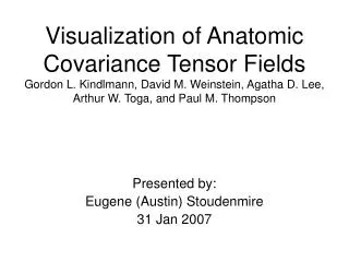
Visualization of Anatomic Covariance Tensor FieldsGordon L. Kindlmann, David M. Weinstein, Agatha D. Lee, Arthur W. Toga, and Paul M. Thompson Presented by: Eugene (Austin) Stoudenmire 31 Jan 2007
Problem • Current methods of visualizing brain variability • Not sufficiently informative and interactive • Current methods involve computing • for each vertex of brain surface model • distribution of displacement vectors between individual brains and average brain • summarizing as covariance tensor • tensor – akin to vector field • And displaying as ellipsoid tensor glyphs • glyph – icon that maps features onto primitive • shape, size, orientation, appearance
Ellipsoidal Tensor Glyphs • But – feature values are hard to discern
We Care • Understanding anatomic variability of the brain is important • Visualization is an important component of understanding anatomic variability
Anatomic Variability Importance • Functional organization differs among people • measures are required to represent and visualize systematic patterns • Determine patterns of altered brain structure in diseases • based on databases of brain scans • Statistical info on anatomical variance to facilitate computer vision algorithms that automatically identify brain structures
Visualization Importance • Important component of understanding anatomic variability • Provide feedback for variability algorithm verification • Means of mucking with the data to form/refine hypotheses about variability
Approach • Map values onto superquadric glyphs • MRI brain scans of 40 subjects • Aligned and converted to 3D models • Interest: Deformation that would transform average onto each individual • Represented as 3D covariance tensor
Ellipsoid Superquadric
Ellipsoid Superquadric Kindlmann, “Superquadric Tensor Glyphs”, Joint Eurographics – IEEE TCVG Symposium on Visualization 2004
Subject Mapping • MRI brain scans of 40 subjects • Aligned to standardized brain • Converted aligned brains to 3D models • Matched up major fissures • Interest: Minimum-energy 3D nonlinear elastic deformation that transforms the landmarks of average onto those of each individual • Represented at each point as 3D covariance tensor of the displacement vectors induced by the deformations from the average to all individuals
Tensor Creation • 3 x 3 covariance tensor T • Diagonalized • T = RR-1 • Where = Diagonal matrix of eigenvalues R = Rotational matrix that transforms standard basis onto eigenvector basis
Tensor Creation • Tensor orientation – Eigenvectors • Tensor shape – Eigenvalues Kindlmann, “Superquadric Tensor Glyphs”, Joint Eurographics – IEEE TCVG Symposium on Visualization 2004
What Makes Superquadric So Good • Edges of superquadric tensor glyph • indicate eigenvalue differences • Eigenvalue differences • imply lack of rotational symmetry • Therefore need to emphasize glyph edges • which will emphasize eigenvalue differences
Tensor Creation • Create superquadric shape • Modify standard parameterization of sphere cos sin sin sin cos ) ( 0 <= <=2 p() = , 0 <= <= where x = sgn(x)|x|
And More • Colormaps were used • To make large-scale patterns stand out • Depicted two tensor attributes simulataneously • Different map on mesh surface and glyph • Frobenius norm (overall variability) • Fractional anisotropy (extent to which variability extends more in some directions than others (e.g. not rotationally symmetrical)) • Skew (precise manner of difference from a sphere)
Evaluation • Back to the beginning, importance was • Understanding anatomic variability of the brain • Visualizing in order to understand anatomic variability • Comparative images were used for verification that the visualization was more discriminatory than elliptical tensors • These pictures, along with those of other references, appeared to more clearly differentiate among attribute values than did ellipsoid glyphs • No objective verification or validation • But • Their previous work referenced studies that did quantitatively measure an increased discrimination when superquadratic tensors were used.
Ellipsoid Superquadric Kindlmann, “Superquadric Tensor Glyphs”, Joint Eurographics – IEEE TCVG Symposium on Visualization 2004
Ellipsoid Superquadric Kindlmann, “Superquadric Tensor Glyphs”, Joint Eurographics – IEEE TCVG Symposium on Visualization 2004
Ellipsoid Superquadric Scientific Computing and Imaging Institute website
Alternative Methods • Spinor mentioned in another paper • Other methods from “Tensorlines: Advection-Diffusion based Propagation through Diffusion Tensor Fields”, David Weinstein, Gordon Kindlmann, Eric Lundberg • Brush strokes (stroke shape, color, texture) • Glyphs • Ellipsoids (one kind of glyph) • Stream-polygons (show info along a path) • Hyperstreamlines (show info along a path) • Other work of theirs did discuss ray-tracing the resulting superquadratic tensors with the caveat that it wouldn’t be real time
Conclusion • Computational efficiency – could be interactive, depending on problem size • Not designed to definitively depict attribute value • Certainly not designed as a physician’s diagnostic tool • Could be applicable to many other uses • Example calculation sure would have been helpful!
Next Steps • Other applications • Efficiency • Additional attributes (e.g. their colormapping scheme)
Question • What are the advantages of superquadric tensor fields (or are there any)
Question • Do the shapes really convey the info they are supposed to (i.e. differences)