Retinal Arterial Macroaneurysm: Case Presentation and Clinical Management Insights
This case presentation discusses an 83-year-old female patient experiencing blurred vision in her left eye for two weeks, accompanied by decreased visual acuity and localized intraretinal hemorrhage. A comprehensive ocular examination revealed findings consistent with cystoid macular edema (CME) and a retinal arterial macroaneurysm. The presentation reviews the potential complications, including hemorrhage and edema, and outlines management options ranging from observation to laser treatments and intravitreal injections. This highlights the importance of proper diagnostic imaging and tailored therapeutic strategies in retinal vascular diseases.

Retinal Arterial Macroaneurysm: Case Presentation and Clinical Management Insights
E N D
Presentation Transcript
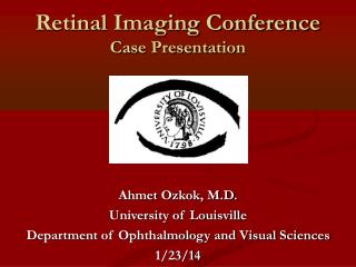
Retinal Imaging Conference Case Presentation Ahmet Ozkok, M.D. University of Louisville Department of Ophthalmology and Visual Sciences 1/23/14
History • CC: ‘blurred vision inthe left eyefor 2 weeks’ • HPI: 83 y/o C F;Blurred visionand trouble focusing noticed 2 weeks ago, gradually increasing. • PMHx: Hyperlipidemia, Arrhythmia, HT • Meds: ASA, Simvastatin, lisinopril • Allergies: NKDA • Family Hx: Heart disease
4→2 20/20 (pl) 4→2 • 20/50 (+0.25, +0.25×015) Ocular Examination VAcc P No RAPD OU EOM: full OU CVF: full OU IOP: 17 / 13 mmHg Anterior Segment : WNL OU except for PCIOL (OU)
FA 00:15 00:21 00:24 D 00:29 00.52 05:21
FA 171 µ
Assessment • Assessment: 83 y/o female with decreased vision, localized intraretinalhemorrhage in the macula, CME, and aneurysm in the left eye.
Differential Diagnosis • Retinal arterial macroaneurysm • Branch retinal vein occlusion
Impression • CME associated with retinal arterial macroaneurysm
Course 28th month 20/40
Retinal Arterial Macroaneurysm • 1/9000 in Beijing eye study • 90% unilateral • Most commonly develop in women (70%/30%) aged between 50-80 • Often associated with HT (65%)
Retinal Arterial Macroaneurysm • Saccular or fusiform dilations of the retinal arterioles within the first three orders of arteriolar bifurcation • They are usually located at the side of an arteriolar bifurcation or AV crossing • ST artery is the most commonly reported site.
Retinal Arterial MacroaneurysmPresentation • Presents with decline in visual acuity due to retinal edema, exudation or hemorrhage • Hemorrhagic RAM • Vitreous, sub-ILM, intraretinal, subretinal, sub-RPE • Exudative RAM • Quiescent RAM
Retinal Arterial MacroaneurysmImaging • FA for demonstration of RAM • ICG may be helpful in cases with dense hemorrhage • Anatomical nature of aneurysm • Saccular • Fusiform Eye (2006) 20, 1011–1020
Retinal Arterial Macroaneurysm • Fibrotic changes in the media of arterial wall • Traction on the adventitia of that segment both by medial layer and ILM • Z-Shaped kink sign
Retinal Arterial MacroaneurysmManagement • Spontaneous obliteration of aneurysm with functional recovery is possible • Aneurysm • Direct photocoagulation to the aneurysm itself • Hemorrhage • Pneumatic displacement w/wotPA • Yag laser for premacular hemorrhage (Photodistruption of ILM) • Vitrectomy
Retinal Arterial MacroaneurysmManagement • Macular edema • Observe • Focal/grid laser • IVTA • Anti-VEGFs
Retinal Arterial MacroaneurysmManagement Am J Ophthalmol 2013;155:898–904
Conclusion • RAM is a rareretinalvasculardisease • Commonlyassociatedwith HT • Can causedecreasedvision • Macularedema • Hemorrhage • FA or ICG is helpfulfordiagnosis • Complicationorientedtheraphy
Ryan, Stephen J.; Schachat, Andrew P.; Wilkinson, Charles P.; Hinton, David R.; Sadda, SriniVas R.; Wiedemann, Peter (2012-11-01). Retina (Ryan, Retina) (Kindle Location 60653). Elsevier Health Sciences. Kindle Edition. • Cho HJ, Rhee TK, Kim HS, et al. Intravitrealbevacizumabforsymptomaticretinalarterymacroaneurysm. Am J Ophthalmol 2013;155:898-904 • Moosavi RA, Fong KC, Chopdar A. Retinalarterymacroaneurysms: clinicalandfluoresceinangiographicfeatures in 34patients.Eye (Lond). 2006 Sep;20(9):1011-20. Epub 2005 Sep 2. • .