Download
1 / 8
80 likes | 287 Vues
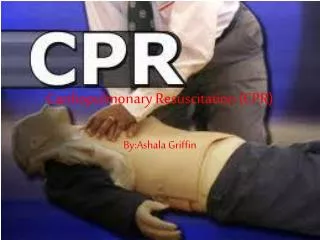
Basic CPR competency is a foudational skill in both basic and advanced life support training and ample data supports the need to improve ongoing maintenance of competency. Many out of hospital cardiac arrest victims do not receive CPR before the arrival of professional rescuers. Video based instruction effectively trains students more quickly than traditional classroom based courses and evidence suggests ongoing refresher training benefits skill retention. Real time feedback devices improve CPR quality in both training and actual resuscitation. Devkunwar Salam "Cardiopulmonary Resuscitation" Published in International Journal of Trend in Scientific Research and Development (ijtsrd), ISSN: 2456-6470, Volume-3 | Issue-2 , February 2019, URL: https://www.ijtsrd.com/papers/ijtsrd21417.pdf Paper URL: https://www.ijtsrd.com/other-scientific-research-area/other/21417/cardiopulmonary-resuscitation/devkunwar-salam<br>

E N D
International Journal of Trend in Scientific Research and Development (IJTSRD) Volume: 3 | Issue: 2 | Jan-Feb 2019 Available Online: www.ijtsrd.com e-ISSN: 2456 - 6470 Cardiopulmonary Resuscitation Devkunwar Salam B.SC Dialysis Technology, Pt. J.N.M Medical Collage,Raipur, Chhattisgarh, India ABSTRACT Basic CPR competency is a foudational skill in both basic and advanced life support training and ample data supports the need to improve ongoing maintenance of competency. Many out-of-hospital cardiac arrest victims do not receive CPR before the arrival of professional rescuers. Video-based instruction effectively trains students more quickly than traditional classroom based courses and evidence suggests ongoing refresher training benefits skill retention. Real time feedback devices improve CPR quality in both training and actual resuscitation. Keywords: CPR, ADVANCE LIFE SUPPORT BASIC LIFE SUPPORT INTRODUCTION CPR is a technique involving heart and lungs that is used when breathing stops. Administering CPR can restore breathing and restart the heart if heart failure accompanies the loss of breathing. This valuable technique should be learned by all caregivers and parents in case an emergency arises where professional help is not immediately available. Emergencies happen all too often, and early intervention can save a life. This lesson will cover how to secure the scene of an accident to protect the victim and the first responders. ?It is a sequence of techniques used to sustain life in the absence of spontaneous breathing and heart beat ?Together chest compressions and rescue breath are called cardiopulmonary resuscitation ?The aim of CPR is to maintain victim's breathing and circulation until emergency aid arrives. CAUSES OF CARDIAC ARREST:- 1.Hypoxia 2.Hypotension 3.Hypoglycimia 4.Acidosis 5.Hypokalemia electrolytes embalance 1. cardiac tamponade 2. tension pneumothorax 3. toxicity (digoxin, local infection) CPR IS WORKS The air we breathe in travels to our lung where oxygen is picked up our by blood and then pumped by the heart to our tissue and organs, when a person experiences cardiac arrest whether due to heart failure in adults and the elderly or an injury such as near drowning, electrocution or severe trauma in a child-the heart goes from a normal beat to an arrhythmic pattern called ventricular fibrillation, and eventually ceases to beat altogether. This prevents oxygen from circulation throughout the body, rapidly killing cells and tissue. In essence, cardio (heart) pulmonary (lung) resuscitation (revive, revitalize) serves as artificial heartbeat and an artificial respirator. DIAGNOSIS OF CARDIAC ARREST- 1.loss of consciousness 2.loss of apical & central pulsations (carotid, femoral) 3.apnea CPR may not save the victims even when performed properly, but if started within 4 minutes of cardiac arrest and defibrillation is provided within 10 minutes, a person has a 40% chance of survival. TYPES (FORM) OF CARDIAC ARREST:- 1.Asystole (isoelectoric line) 2.Ventricular fibrillation (VF) 3.Pulseless ventricular trachycardia (VT) 4.pulseless electrical activity (PEA) @ IJTSRD | Unique Reference Paper ID – IJTSRD21417 | Volume – 3 | Issue – 2 | Jan-Feb 2019 Page: 655
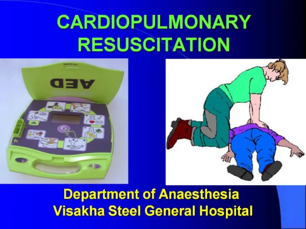
International Journal of Trend in Scientific Research and Development (IJTSRD) @ www.ijtsrd.com eISSN: 2456-6470 SRART CPR IMIDEATEIALY:- ?SEQUENSES OF CPR:- ?Check responsiveness ?Call for help ?Correctly place the victim and ensure the open airway ?Check the presence of spontaneous respiration ?Check pulse ?Start CPR CPR IS TWO MAIN STAGES 1.Basic life support (BLS) :- 2.Advanced life support (ACLS):- Basic life support (BLS) :-It is life support without the use of special equipment Advanced life support (ACLS):- it is life support with the use of special equipment (eg. Airway, endotracheal tube, defibrillator). -3 steps before the initiation of resuscitation for management of a collapsed patient 1.ensure your own safety 2.check the level of responsiveness by gently shaking the patients and shouting: “are you alright?” 3.shout for help. ?Then check for carotid pulsation. ?Apnea (cessation of respiration) is conformed by:- 1.Look: to see chest wall movement. seesaw (paradoxical) movement of the chest wall indicates airway obstruction. 2.listen: to breath sounds from the mouth. 3.feel: air flow for at least 10 seconds. ?Refers to the period of time immediately following trauma during which approximately 50% death occurs. ?The causes of death is usually preventable provided that adequate resuscitation, intervention are provided. (eg. Tension penumothorax , cardiac temponade). LIFE SUPPORT INCLUDES C-A-B C= Circulation. A= Airway. B= Breathing C = Circulation (A)CHEST COMPRESSION – (Cardiac massage ) The human brain cannot survive more then 3 minutes with lack of circulation. So chest compression must be started immediately for any patients with absent central pulsation. Techniques of chest compression: 1.patients must be placed on a hard surface (wooden board). 2.The palm of one hand is placed in the concavity of the lower half of the sternum 2 fingers above the xiphoid process. (Avoid xiphisternal junction –fracture & injury ) ?The other hand is placed over the hand on the sternum. ?Shoulder should be positioned directly over the hands with the elbows looked staright and arms extended. ?Sternum must be depressed 4-5 cm in adults. and 2-4 cm in children, 1-2 cm in infants. ?Must be performed at a rate of 100 – 120/min. ?During CPR the ratio of chest compressions to ventilation should be as follows. ?Single rescuer = 30:2 ?In the presence of 2 rescuer chest compression must not be interrupted for ventilation. CHEST OF COMPRSSION IN INFANTS (0-12 MONTHS) Check for Responsiveness - 1.Check for responsiveness Shake or tap the child gently. See if the child moves or makes a noise. Shout, "Are you okay? “ 2.If there is no response, shout for help. Send someone to call 911 and retrieve an automated external defibrillator (AED) if one is available. Do not leave the child alone to call 911 and retrieve an AED until you have performed CPR for about 2 minutes 3.Carefully place the child on his or her back. If there is a chance the child has a spinal injury, two people should move the child to prevent the head and neck from twisting diagnosis and surgical @ IJTSRD | Unique Reference Paper ID – IJTSRD21417 | Volume – 3 | Issue – 2 | Jan-Feb 2019 Page: 656
International Journal of Trend in Scientific Research and Development (IJTSRD) @ www.ijtsrd.com eISSN: 2456-6470 Chest Compression- 4.Perform chest compressions ?Place the heel of one hand on the breastbone -- just below the nipples. Make sure your heel is not at the very end of the breastbone. ?Keep your other hand on the child's forehead, keeping the head tilted back. ?Press down on the child's chest so that it compresses about 1/3 to 1/2 the depth of the chest. Give 30 chest compressions. Each time, let the chest rise completely. These compressions should be FAST and hard with no pausing. Count the 30 compressions quickly. Child Not Breath- 5. Open the airway. Lift up the chin with one hand. At the same time, push down on the forehead with the other hand. 6. Look, listen and feel for breathing. Place your ear close to the child's mouth and nose. Watch for chest movement. Feel for breath on your cheek. 7.If the child is not breathing: ?Cover the child's mouth tightly with your mouth. ?Pinch the nose closed. ?Keep the chin lifted and head tilted. ?Give two breaths. Each breath should take about a second and make the chest rise. 8. Continue CPR (30 chest compressions followed by 2 breaths, then repeat) for about 2 minutes. 9. After about 2 minutes of CPR, if the child still does not have normal breathing, coughing, or any movement, leave the child if you are alone and call 911. If an AED for children is available, use it now. 10. Repeat rescue breathing and chest compressions until the child recovers or help arrives. If the child starts breathing again, place him or her in the recovery position. Periodically re-check for breathing until help arrives. COMPILCATIONS OF CHEST COMPRESSION: ?Fractured ribs (most commonly). ?Pneumothorax. ?Sternal fracture. ?Anterior mediasitnal hemmorage. ?Injury to abdominal viscera (eg. Liver) ?Pulmonary complication (contusion). ?Rarely injury to heart and great vessels (eg. Myocardial contusion) (very rerly). ?Usually AVOIDABLE by perfoming the technique correctly. ?Chest compression must be continued for 2 minutes before reassessment of cardiac rhythm. (2 minutes = equivalent to 5 cycles 30:2 ) ?Golden rules :- • Ensure high quality chest compression :rate depth,recoil. • Plan action before interrupting CPR. • Minimize interruption of chest compression. • Early defibrillation of chest compressions. Early defibrillation of shockable rythm. @ IJTSRD | Unique Reference Paper ID – IJTSRD21417 | Volume – 3 | Issue – 2 | Jan-Feb 2019 Page: 657
International Journal of Trend in Scientific Research and Development (IJTSRD) @ www.ijtsrd.com eISSN: 2456-6470 (B) Defibrillation: Adult ALS algorithm. VENTRICULAR TRCHYCARDIA (VT) SHOCKABLE – ?Broad bizarre-shaped complex. ?Rapid rate :120-125/min ?Regular ?Precordial thumb: rapid treatment of a witneesed and monitored VF/VT cardiac arrest. ASYSTOLE (NON-SHOCKABLE) ?Check that all lead are attached ?Adrenaline 1 mg IV every 4 mins ( 2 cycles ) (until a shockable rhythm is achieved). PULSELESS ELECTICAL ACTIVITY (PEA) NON SHOCKABLE ?Exclude / treat reversible causes. ?Adrenaline 1 mg iv every 4 mins (2 cycles ) (until a shockable rhythm is reached). VENTRICULAR FIBRILLATION (VF) SHOCKABLE – ?Bizarre irregular waveform. ?No recognizable QRS complexes. ?Random frequency and amplitude. ?Coarse / fine ?Exclude artifact.-1 movement 2electrical interference DEFIBRILLATION TECHNIQUE -: Position of paddles – ?One paddles is placed in the right infraclavicular region, while the other is placed in left 5th 6th intercostal space anterior axillary line. ? Alternatively antero – posterior may be used : one paddles is placed in the infrascapular region while other is placed in the left 5th- 6th intercostal space anterior axillary line. @ IJTSRD | Unique Reference Paper ID – IJTSRD21417 | Volume – 3 | Issue – 2 | Jan-Feb 2019 Page: 658
International Journal of Trend in Scientific Research and Development (IJTSRD) @ www.ijtsrd.com eISSN: 2456-6470 ?consider in defibrillation station ?safety first – teamwork vital ?wording / exact language used is less important than communicating principles to ensure safety. ?Not a script. ?Chest compressions while defibrillator charging ?first time rhythm checked is when compressions are ceased. ?Complication of defibrillation: skin burn injury to myocardium and elevation of cardiac enzymes .electrocution of person in contact with bed. A-AIRWAY AIRWAY- loss of consciousness often results in airway obstruction due to loss of tone in the muscles of the airway and falling back of the tongue. (A)-Basic techniques for airway patency:- 1. Head tilt, chin lift:- one hand is placed on the forehead and other on the chin the head is tilted upwords to cause anterior of displacemets of tongue. 2.jaw thrust:- precaution: ?Make sure the paddles have conducting gel on them: ?1-the electricity will not be properly transmitted to the chest wall without it. ?2-even with the gel these paddles will often a second – degree skin burn . ?make sure you have cleared the bed:make sure that no one is in contact with the bed otherwise this person may be electrocuted and develop VT or VF. ?Hold the paddles down firmly. ?Chest compressions must be continued for 2minutes after DC shock before reassessment of cardiac rhytham. 3.Heimlich manoeuvre:- if the pt is conscious or the foreign body cannot be removed by a finger sweep.it is done while the pt is standing up or tying down . this is a subdiaphragmatic abdominal thrust that elevates the diaphragm expelling a blast of air from the lungs displaces the foregion body. In infants his can be done by a series of blowsb on he back and chest thrusts. @ IJTSRD | Unique Reference Paper ID – IJTSRD21417 | Volume – 3 | Issue – 2 | Jan-Feb 2019 Page: 659
International Journal of Trend in Scientific Research and Development (IJTSRD) @ www.ijtsrd.com eISSN: 2456-6470 ?Lubricated and inserted throught the nose. ?Better tolerated in conscious patients. ?Contraindicated :in anticoagulant patients and fractured skull base. ?Disadvantages: not protective against regurgitation & aspiration of gastric contents. 4.laryngeal mask (LMA):- Cervical spines :- 1.special care must be taken during airway management for the cervical spines. Any polytraumatised patient may sustain injury to the cervical spines and any rough manipulation may results in cervical spinal cord injury and subsequent quadriplegia. 2.thus in any polytraumatised patient cervical in line- stabilization must be routinely performed during transport and airway management. 3.this can be done by a cervical collar. 4.the patients should be transported by specially trained medical personnel as one unit. (B) Advanced techniques for airway patency :- 1. Facemask -1 foggy mask. 2. rising chest. ?Available in a variety of pediatric and adults sizes. ?Advantages: easy. does not require highly skilled personnel (can be used by paramedics ). ?Disadvantages: stomach inflation. Not protective against regurgitation & aspiration of gastric content. 5.Endotracheal tube:- Advantages: easy does not require skilled personnel (paramadies). Disadvantages:-stomach inflation .not protective against regurgitation & aspiration of gastric contents. 2. Oropharyngel Airway – Advantages: ensures proper lung ventilation. No gastric inflation. No regurgitation or aspiration of gastric contents. Disadvantages: requires insertion by highly skilled personnel. 6.Combitube:- Advantages: easy .does not require highly skilled personnel (can be used paramedics) . Disadvantges: not protective against regurgitation & aspiration of gastric content .poorly toler ated by conscious patients. 3.Nasopharyngeal Airway- Advantage: easy to use. Does not require highly skilled personnel (can be used by paramedics). @ IJTSRD | Unique Reference Paper ID – IJTSRD21417 | Volume – 3 | Issue – 2 | Jan-Feb 2019 Page: 660
International Journal of Trend in Scientific Research and Development (IJTSRD) @ www.ijtsrd.com eISSN: 2456-6470 7.cricothyrotomy:(surgical airway ). B-BREATHING Breathing with the victims airway clear of any obstruction, gently, support his chin so as to keep it lifted up and the head tilted back, pinch his nose to prevent air from escaping once you begin to ventilate. (A)Basic techniques include:- 1. mouth to mouth breathing:- with the airway held open, pinch the nostrils closed, take a deep beath and seal your lips over he patients mouth. blow steadily into the patients mouth watching the chest rise as if the patients was taking a deep breath. 2.mouth to nose breathing:- seal the mouth shut and breathe steadily though the nose. 3.mouth to mouth and nose:- is used in infants and small children. 4. successful breathing is achieved by delivery of a tidal volume of 800 -1200 ml in adults at a rate of 10-12 breath / min in adults. (B) Advanced techniques include:- 1. Self – inflating resuscitation bag ( Ambu bag ). ?When used without a sourse of O2(room air) gives 21% .O2 . ?When connected to a source of O2 (10-15L/min) gives 45% 02 ?If a reservoir bag is added it can give up to 85% O2. 2.Mechanical ventilator in OR or in ICU. DRUGS USED IN CPR:- Adrenaline:- ?Given as a vasopressor a-1 effect (not as an inotrope). ?Dose:1mg (0.01 mg/kg) Iv every 4 minutes ?(alternating cycles )while continuing CPR. ?Given: 1.immediately in non – shockable rhythm (non- VT/VF). 2.in VF or VT given after the 3rd shock. ?Repeated: in alternative cycles (every 4 minutes ). ?Once adrenaline - always adrenaline. Amiodarone:- ?Dose_ 300 mg IV bolus (5 mg/kg). ?Given: in shockable rhythm after the 3rd shock. ?if unavailable give lidocaine 100 mg IV (1-1.5 mg/kg). Vasopressin: - (ADH): 40 mg IU single dose once. Magnesium: - Dose: 2 g IV ?Given: 1.VF/VT with hyomagnesemia 2.Torsade de pointes. 3.Digoxin toxicity. Calcium:- ?Dose: 10 ml of 10% calcium chloride IV. ?Indication: PEA caused by: hyperkalemia, hypocalcemia, hypermagnesemia,and overdose of calcium channel blockers. ?Do not give calcium solutions and NaHCO3 simultaneously by the sme route. ?It is done either by a commercially available cannula in a specialized cricothyrotomy set or a large bore iv cannula 12-14gauge. ?Is done in case of difficult endotracheal intubation. ?Nu- trake cannula is specially desigened to allow ventilation by a self –inflating bag (AMBU) ?An iv cannula needs a special connection to a high pressure source to generate sufficient gas flow ( trans- tracheal jet ventilation). 8.Tracheostomy:- (surgical airway). is a surgical procedure which consist of making an incision the anterior aspect of the neck and opening a direct airway through an incision in the trachea. The resulting stoma or tracheostomy.can serve independently as an airway or as a site for a tracheal tube or tracheostomy tube to be inserted this tube allow a person to breathe without the use of the nose or mouth. @ IJTSRD | Unique Reference Paper ID – IJTSRD21417 | Volume – 3 | Issue – 2 | Jan-Feb 2019 Page: 661
International Journal of Trend in Scientific Research and Development (IJTSRD) @ www.ijtsrd.com eISSN: 2456-6470 IV Fluid:- ?Infuse fluid rapidly if hypovolemia is suspected. ?Use normal saline (0.9% NaCL) or Ringers solution . ?Avoid dextrose which is redistributed away from the intravascular space rapidly and causes hyperglycemia which may worsen neurological outcome after cardiac arrest. ?Dextrose is indicated only if there is documented hypoglycaemia. Avoid its routine use due to its complication: 1.increases Co2 load. 2.inhibitors release of O2 to tissues. 3.impairs myocordial contractility. 4.causes hypernatremia. 5.adrenaline works better in acidic medium. Atropine:- ?Its routine use in PEA and asystole is not beneficial and has become obsolete. ?Indicated in: sinus bradicardia or AV block causing hemodynamic instability. ?Dose: 0.5mg IV. Repeated up to a maximum of 3 mg (full atropinization). Conclusion- Cardiac arrest is an uncommon event during Embrace transport, this audit gave a rate of CPR of 4 per 1000 acute transfers. Collaboration with other transport services to compare this data will improve patient care. By giving CPR we can save life of many people . both basic life support and advance life support system is very important to save many life . REFRANCE:- [1]BLS for health care providers, Student manual .American heart association AHA 2006 Thrombolytics:- ?Fibrinolytic therapy is considerd when cardiac arrest is caused by proven or suspected acute pulmonary embolism. ?If a fibrinolytic drug is used in these circumstances consider performing CPR for at least 60-90 minuts before termination of resuscitation attempts. ?Eg :Alteplase ,Tenecteplase steptokinase ). Sodium bicarbonate:- ?Used in:- 1.severe metabolic acidosis (pH < 7.1 ) 2.life- threatening hyperkalemia. 3.Tricyclic antidepressant overdose. ?Dose : ( half correction ) ½ base deficit ×1/3 body weight. ( Old generation [2]Fundamentals Of Nursing Procedures Book Second year Nursing, 2008. @ IJTSRD | Unique Reference Paper ID – IJTSRD21417 | Volume – 3 | Issue – 2 | Jan-Feb 2019 Page: 662