Vitamins
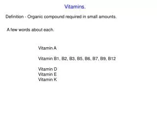
Vitamins. Definitions:. Vitamins are organic compounds required by the body in trace amounts to perform specific function, and can not be synthesized by humans, or can not be synthesized in adequate (sufficient) quantities to meet needs.

Vitamins
E N D
Presentation Transcript
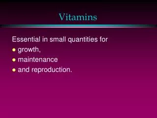
Definitions: • Vitamins are organic compounds required by the body in trace amounts to perform specific function, and can not be synthesized by humans, or can not be synthesized in adequate (sufficient) quantities to meet needs. • When present in inadequate quantities, deficiency states results leading to disease. • Modern views suggest that more quantities than RDIS might be needed to prevent some chronic diseases.
Definitions: • Provitaminsare precursors of vitamins that could be converted into vitamins inside the body e.g. Carotenes are provitamin A. • Vitamers:These are different forms of one vitamin e.g. Vitamin D has 2 vitamers; D2 &D3.
Common features of fat soluble vitamins: • Released, absorbed and transported with the fat in the diet. • Not readily excreted in the urine. • Excess of immediate need is stored in liver or adipose tissues. • Some cause toxicity in excess.
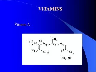
. Vitamin A (retenoids) Carotenes are the provitamin A. The different forms (vitamers) of vitamin A used by the body are :retinol, retinal and retinoic acid
Sources: Liver, kidney, eggs milk fat and fish liver oil. It is also present in yellow and dark green vegetables as provitamin A Carotenes (α, β and γ). Requirements: it is expressed as retinol equivalent where 1 RE equals 1 μg of retinol. RE equals 5 IU and the daily requirement is about 5000 lU.
Absorption and Metabolism • In the intestine, β-carotene is converted into retinal. Retinyl esters also are hydrolyzed and retinol is absorbed. • In the intestinal mucosal cells, retinol is re-esterified with palmitic acid and transported in chylomicrons to the liver, where 90% of the body's vitamin A is stored. • Retinoic acid is absorbed directly into the portal circulation to the liver. • When the body cells need vitamin A, retinyl esters are hydrolyzed and free retinol combines with a protein formed by the liver called retinol binding protein (RBP). • RBP carries retinol to the target cells. When retinol becomes inside the cells, it is bound by cellular retinol binding protein (CRBP).
Functions of vitamin A: • Vision: The human retina contains two types of receptor cells for vision; cones and rods. Vitamin A is a component of visual pigments present in cones and rods. Cone cells are responsible for day vision and color. Rod cells are responsible for vision in poor light e.g. at night.
Visual cycle: • Rhodopsin (the visual pigment of rod cells)consists of protein called opsin bound to 11-cis retinal (double bond at position 11 is in cis form, while other double bonds are in trans form). • When rhodopsin is exposed to light, 11 cis retinal is converted into all trans retinal (all double bonds are in trans form), and dissociates from opsin. • All trans retinal changes the permeability of cell membrane of rod cells. This allows the calcium ions to pass out of the cell membrane. This stimulates the nerve impulse in optic nerve. Thus the brain perceives light. • Rhodopsin must regenerated for vision. All trans retinal is converted back to 11-cis retinal, but this conversion is incomplete. This can be supplied from dietary retinol, which is oxidized to give 11 cis retinal.
Functions of vitamin A (continue) • Vitamin A acts as a hormone. It binds to nuclear proteins and acts on certain genes (by affecting their transcription rate). It has the following functions: • Reproduction: Retinol and retinal are essential for reproduction. They support sperm formation (spermatogenesis) in males and maintain fetal life in females. • Growth: Vitamin A is essential for normal growth and bone formation.
Functions of vitamin A(continue) • Glycoprotein synthesis, and phospholipids synthesisin the lungs (lung surfactant) and for cell differentiation . • Maintenance of epithelial cells: It is essential for normal differentiation of epithelial cells. This is important for smoothness of skin and mucus membranes. • Antioxidant (anticancer) action: Retinoids and carotinoids (Carotenes) act as antioxidants and protect tissues from toxic effect of some oxidants that may lead to epithelial tissue cancer
Deficiency of vitamin A: 1- Eye: deficiency will lead to night blindness (impaired dark adaptation) and Xero-ophthalmia (dryness and roughness of cornea) and keratomalicia.. 2-Growth retardation.
3-Skin and mucus membranes: roughness of skin and mucus membranes of different body systems e.g. urinary system. This leads to increased susceptibility to infection. • 4-Degeneration of testes and abortion. • 5-Hydrocephalus and increased intracranial pressure
Treatment of deficiency: the usual line of treatment is 220 mg of retinol orally divided on two days. Evaluation of nutritional status:retinol level in serum is the most widely used method, the normal range for children is 20-90 ug/dl, in adult it is 30-90 ug/dl, serum level higher than 100 ug/dl is an indication of toxicity.
Excess vitamin A intake (hypervitaminosis A): • intake of more than 100000 IU/day: Occurs when excessive vitamin A intake exceeds the capacity of RBP. • Free retinol will be released in blood with the following toxic effects; Headache, nausea, bone pain due to bony depositions (hyperostosis) and loss of hair, dermatitis and skin itching. • Vitamin A is teratogenic in high doses (> 15000 IU/day) if taken during the first two months of pregnancy. It may cause congenital abnormalities in the fetus including microcephaly, dilated ventricles and abortion.
RDI: 10 µg/day 400 lU/day
The synthesis of provitamin D3 and Vitamin D3 are self-controlled processes since further absorption of UV light causes isomerizations of these compounds to yield inactive products. • In human system, the production of Vitamin D3 in skin is important, since nutritional supply of Vitamin D3 as well as Vitamin D2 is limited. • the sources of dietary vitamin D are limited primarily to liver, eggs, butter, fortified milk and fatty fish. • Conversion of 7-dehydrocholesterol to previtamin D3 is decreased to less than half in elderly people. • From skin Vitamin D3 enters to circulation where all Vitamin D compounds are mainly bound to Vitamin D binding protein (DBP). • Dietary Vitamin D2 and D3 enter the circulation in the chylomicrons. • In the body, both Vitamin D2 and D3 undergo similar activation processes, which are prerequisite for their biological activity.
Functions of vitamin D • Vitamin D plays an important role in calcium and phosphate homeostasis • It helps to increase serum calcium and phosphate by: 1- Increasing intestinal absorption. 2- Decreasing renal excretion. 3- Increasing bone resorption.
Functions of vitamin D not related to calcium homeostasis: • Beside its very well known function in calcium and phosphate metabolism ,Vit. D receptors are found in many tissues , thus it can exert its effects on them.Examples: A- Vit. D stimulate synthesis of non-collagenous bone matrix proteins, e.g. osteocalcin ,osteopontin B- It is possibly needed for the regulation of differentiation and proliferation of various cells including immuno regulatory cells, epidermal cells and malignant tumor cells.
Functions of vitamin D not related to calcium homeostasis (continue): • It was found that vitamin D causes arrest of cell in G1 phase and induces apoptosis. • It promotes monocyte differentiation and inhibits lymphocyte proliferation and secretion of cytokines, such as IL-2, interferon-γ and IL-12. • Vit. D analogues are tried therapeutically eg. treatment of psoriasis (vit D induces differentiation and inhibits proliferation in keratinocytes).
Functions of vitamin D not related to calcium homeostasis (continue): • In several different types of cancer cells Vitamin D has been shown to have anti-proliferativeeffects. • Vit D and Ca deficiency are associated with incidence of colon and breast cancers. • In vitro studies show vitamin D analogues inhibit the proliferation of these cells.
Deficiency of vitamin D: • Causes demineralization of bones, resulting in rickets in children and osteomalaciain adults , and is usually characterized by low serum calcium and phosphate, and increased alkaline phosphatase and parathyroid hormone levels. • Ricketsis characterized by the continued formation of the collagen matrix of bone , but incomplete mineralization, resulting in soft , pliable bones.
In osteomalacia demineralization of preexisting bones increases their susceptibility to fracture. • Inability to activate to activate the vitamin in chronic renal failure, results in renal rickets( renal osteodystrophy)
Vitamin D Nutritional Status • Vitamin D nutritional status can be evaluated by measuring plasma levels of 25 OH D3 and 1 α,25 (OH)2 D3. • Serum level of 25 OH D3 represent an index of vitamin store, while 1 α,25 (OH)2 D3 is less reliable. • The acceptable level for adult is 20 ng/ml for 25 OH D3 and 20-40 pg/ml for 1 α,25 (OH)2 D3. • Certain drugs as anticonvulsants used for long time may lead to rickets or osteomalacia.
Excess vitamin D (overdose or hypervitaminosis D): • The most toxic of the vitamins. • High doses can cause loss of appetite , nausea, thirst, and stupor. • Also causes increased calcium absorption and bone resorption resulting in hypercalceamia. • This leads to abnormal calcification of tissues and deposition of calcium and phosphate in different systems e.g. urinary system causing stone formation.
Vitamin E (tocopherols) Structure: there are four types of tocopherols α , β, γ and δ. All of them contain tocol ring. The most active form is α tocopherol (as antioxidant). , β, γ and δ tocopherols differ from α tocnopherol in number and position of - CH3 groups attached to the tocol ring.
Sources: • green leafy plants and seed oils are the best sources. The richest sources for human are salad oils, margarines derived from soybean, peanut, corn and safflower oils. Animal sources containing the highest amount include eggs, liver, and muscle meats. Requirements: 11-15 mg/day
Absorption and Transport • absorption requires bile salts and pancreatic esterase enzyme. • The vitamin is incorporated in the chylomicrons. • Most of the absorbed vitamin is deposited initially in the liver and then distributed to the adipose tissues. • In serum, two thirds of the vitamin is transported bound to LDL with the rest carried by the other lipoproteins. • In both the cardiac and hepatocyte cytoplasm there is specific carrier protein that carries the vitamin into the mitochondria.
Function of Vitamin E • vitamin E acts as antioxidant; it prevents non-enzymatic oxidation of cell components (e.g. polyunsaturated fatty acids, DNA and cell membranes) by molecular oxygen or free radicals. By this function vitamin E can prevent or decrease the oxidation of LDL, and maintain membrane integrity. • Therefore ,The vitamin is found in all cell membranes, the greatest amount is found in adipose tissue.
Deficiency:Occurs usually in premature infant, in people with fat malabsorption or biliary obstruction. Deficiency leads to hemolysis of RBCs and anemia: due to lack of protection against peroxides and also leads to muscle breakdown. • It was found that people with cardiovascular diseases and cancer have lower levels of serum vitamin E compared to the healthy people.
Signs, symptoms and treatment of deficiency: • they are not specific including hemolytic anemia, myopathy, weakness, ataxia, impaired reflexes, ophthalmoplegia, retinopathy. In severe deficiency, permanent damage to nerve tissues takes place. • Deficiency states may be corrected with oral intake of 0.2-2.0 g/day.
Toxicity • Toxicityof vitamin E is not observed up to more than 2000 mg/day. But doses of more than 100 mg/day can prevent the activation of vitamin K, so it should be avoided during anticoagulant therapy.
Use and effect of large doses: certain rare inborn error of metabolism respond to high doses of vitamin E e.g. glucose-6-phosphate dehydrogenase deficiency, glutathione peroxidase deficiency. • Vitamin E is relatively non toxic even in very high doses. But very high doses for long period may impair immune function and may interfere with arachidonic acid and prostaglandin metabolism.
Evaluation of nutritional status: • the best method is measuring vitamin E level in blood, normal values are 0.5-1.2 mg/dl. • Vitamin E mobilizes from the liver bound to VLDL therefore hypolipidemic patients will have lower serum levels. • There is some evidence that a ratio of serum tocopherol to total lipid is a better indicator of nutritional state. • A value below 0.8 mg tocopherol/g of total serum lipid is considered deficient in adult and children
Vitamin K • Structure:There are three forms (vitamers) of vitamin K: K1, k2 and K3 The difference between KI and k2 lies in the side chain R. K3 is synthetic vitamin and has no R side chain.
Sources: • The main source of vitamin K is the intestinal bacteria. They produce Vitamin K2 (menaquinones). • Vitamin K1 (phylloquinones) is of plant origin (green leafy vegetables, olive, soybean and cauliflower) • It is also found in egg yolk and liver. • Vitamin K3 is synthetic, , water soluble and more potent than vitamin K1 and K2.
Absorption and storage:vitamin K is absorbed from the intestine in the chylomicrons , and transported to the liver, and from liver to the tissues incorporated in VLDL. High doses of other fat soluble vitamins decrease vitamin K absorption. • Tissue store of the vitamin is less than the store of other fat soluble vitamins. Vitamin K localizes in various cellular membranes, particularly in the golgi and smooth microsomal membrane fractions. • Vitamin K1 is rapidly metabolized to more polar metabolites and excreted in the urine and bile.