Development of a New Endorectal PET-TOF Probe for MRI-Compatible Prostate Cancer Imaging
10 likes | 135 Vues
This project aimed to design and test an innovative endorectal PET-TOF probe that is compatible with MRI and MRS for effective diagnosis and follow-up of prostate cancer. Prostate cancer is prevalent in Western countries, making accurate imaging essential. Current imaging techniques like CT and MRI are hindered by spatial resolution issues. Our probe utilizes Silicon Photo Multipliers (SiPM) to enhance sensitivity and resolution, facilitating simultaneous PET-MRI scans. Preliminary studies indicate promising performance, offering hope for improved prostate imaging and diagnosis.

Development of a New Endorectal PET-TOF Probe for MRI-Compatible Prostate Cancer Imaging
E N D
Presentation Transcript
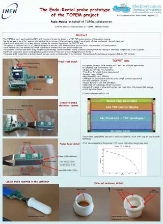
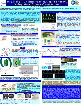
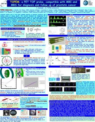
TOPEM: a PET TOF probe, compatible with MRI and MRS for diagnosis and follow up of prostate cancer F. Garibaldi1,, for TOPEM collaboration TOPEM Collaboration: A. Gabrielli, F. M. Giorgi - INFN, University of Bologna - F. Garibaldi, F. Cusanno, F. Meddi, INFN Roma1 and gruppo collegato ISS, B. Maraviglia F. Giove, T. Gigli, University of Roma - R. De Leo, M. Foresta, A. Ranieri, G. De Robertis, F. Loddo - INFN, University of Bari - R. Fonte - INFN, Catania - P. Musico, C. Calvini, P. Ottonello – INFN, University of Genova - L. G. Cosentino, A. D. Pappalardo, P. Finocchiaro – INFN, University of Catania, LNS – N. Clinthorne, S. Huh - University of Michigan, S. Majewsky – University of West Virginia Prostate cancer (PC) is the most common disease in western countries and a leading cause of cancer death. The current standard for diagnosing PC is transrectal ultrasound (TRUS) guided sextant biopsy. It is the only case of blind biopsy. Powerful techniques and instruments such as CT, MRI, PET/SPECT suffer for limited spatial resolution, sensitivity and/or specificity. Dedicated detectors and techniques are needed. Multimodality imaging can play a significant role, merging anatomical and functional details coming from simultaneous PET and MRI (and MRS) scans. A new research project started with the goal of designing, building and testing an endorectal PET-TOF probe compatible with MRI. SiPM Dimension, compatibility with magnetic field, and timing dictate the choice of the photodetector: Silicon Photo Multipliers (SiPM). We started studying the performance of SiPMs having different characteristics (microcell dimension, pixel size etc.) from different companies. The adjacent figures show, as expected, gain reduction with temperature is due to the variation of breakdown voltage. This puts constraints on the layout with need of cooling, monitoring the temperature and feedback on the bias voltage. Preliminary calculations show that by water cooling this is possible. The figure shows a possible layout. Drawback of the standard PET • -- detectors far away from prostate • -poor spatial resolution (6 – 12 mm) • poor photon detection efficiency (<1%) • activity ouside the organ • -> poor contrast resolution A dedicated external PET detector (W.Moses) will improve the performances but it is still limited (by attenuation etc.) Electronics Due to geometrical and technical constraints we will have a detector unit (with ASIC) and a control unit.The detector will have dual layer of scintillators and SiPM (for Depth Of Interaction (DOI) precision measurement). ASIC: minimal implementation will have preamp-discriminators. In this scenario very high density cabling will be necessary for off-detector signal processing. Our goal is to design a final ASIC with all funcionalities (ADC + TDC) with minimal wiring (serial communication). Concept of a dedicated prostate imager An endorectal probe and a PET panel imaging module • Imaging (“zoom”) probe: 1.5 mm LYSO arrays coupled to a MPPC array 2) Panel PET imager: 150 x 200 mm2 LYSO array cuupled to 4x3 H8500 Hamamatsu for testing pourposes. Array of SiPM possibly coupled to LYSO (or LSO array doped with Ce). TOF measurement or We performed preliminary measurement of timing resolution with a finger LYSO scintillator obtaining ~ 350 ps FWHM. Substantial improvements are possible showing that the design goal (≤ 300 ps) is reachable Using an external standard PET detector and 1 or 2 panel detectors. High resolution possible if internal detector has high resolution Attenuation of activity emitted from prostate lower. (N. Clinthorne et al.). External high resolution detectors can augment imaging of superficial lymph nodes. Measurements with test detector(s) Geant4 simulations using Zubal antropomorphic phantom -Model 80cm x 15cm x 2cm BGO external ring -Internal LSO probe, 2 layers of 1mm x 1mm x 3mm crystals in a 9 x 35 array -source in prostate 20mm from probe face z The panel detector: an array of 4x3 Hamamatsu flat panel H8500 PMTs was coupled to the Saint Gobain array of 2x2x15mm LYSO scintillator pixels, to form the active FOV imaging surface of about 15cm x 20cm. . x Probe: (1 x 1 x 10 mm2) Lyso. One side readout with Hamamatsu SiPM Laminography: thirteen planes spaced at 2.5mm in the z-directionand covering z region between -15mm to +15mm.“Plus” direction is away from the panel imager and towards the probe. All reconstruction angles, limited only by the detector sizes and source geometry, were accepted. Each of the three point sources is best seen “in focus” in one of the planes (marked with red arrows), as expected from the 6 mm vertical (z) spacing of the sources. The planar spacing (seen here in the vertical image coordinate) is 5mm. 1.3 mm 0.8 mm 1.2 mm 0.9 mm Spatial resolution and efficiency increase dominated by probe resolution and dimension. Adding one or two panel detectors close to the patient’s body would increase the efficiency helping also detecting the lymph nodes 1.1 mm 1.0 mm Three Na22 point sources spaced vertically at 6mm, and side shifted by 5mm, 45 deg acceptance angle. Advantages of TOF The spatial resolution of the system in the magnified geometry, similar to the one expected in imaging of the prostate, is approaching ~1.5mm FWHM (with 1mm LYSO array) in plane when using simple laminography back projection algorithm.The vertical (z) resolution is on the order of 5-10mm FWHM depending on the imaging geometry • - TOF provides huge performance increase! • Can localize the source along line of flight. • In non TOF case the voxels are coupled, the statistical noise is increased. TOF information reduces the coupling, and reduces statistical noise. • If the distance between 2 voxels> DX=cDT/2 they are uncoupled. • increase SNR(NECR) • - Fast convergence (reduced scan time) Radiotracers: good radiotracers are an issue. Recently PET-Choline and other tracers gave better results than FDG. Moreover, much better tracers will be available soon. With 300 ps FWHM, 4.5 cm (just the dimension of the prostate). This is challenging but possible Summary and Outlook: A project started, with the goal of designing, building, testing on phantoms and possibly on animals (and humans) an endorectal PET-TOF MRI probe to be used in coincidence with an external dedicated detector and/or a standard PET. New, very promising radiotracers will be tested on animals.Using SiPM will allow both good TOF resolution (and consequently great improvement in NECR) and full compatibility with MRI&MRS. Multimodality and improvements in spatial resolution and efficiency will be a crucial step forward in the diagnosis and follow up of the prostate cancer. TOF resolution, MRI compatibility, electronics (mainly the ASIC), miniaturization, algoritmics and software for full integration of the probe with panel detector(s) and a standard PET scanner are the major challenges. Preliminary evaluations, simulation and measurements show that we can build a system fully matching the design performance. Multimodality The advantages of combining PET (functional) with MRI (morphological and even functional). MRS offers an adjunct advantage(inversion of citrate-choline in tumor). This is challenging but it has been shown possible (see B. Pichler et al. Nat. Med.Vol 14, N 4, April 2008) Reference: s1.G. Kellof et al. Challenges in Clincal Prostate Cancer: Role of Imagin. AJR:192, June 2009, pg. 1455. M. Pomper et al. New Agents and Techniques for Imaging Prostate Cancer. Focus on molecular Imaging. To be published in JNM. 2. S.S Huh, N. Clinthorne, W.L. Rogers. Investigation of an internal PET probe for prostate cancer Imaging, NIMA 579 (2007 339-343 3. Tay YC et al. Initial study of an asymmetric PET System dedicated to breast cancer imaging. IEEE TNS 53: 121-126 4. N. Clinthorne et al. Multi-resolution image reconstruction for high resolution small anima PET device. IEEE 2003, Nucl Symp. Conf. Rec. 3: 1997-2001 5. J.sS Karp et al. Benefit of Time –of-Flight in PET: Experimental and Clinical Resukts, JNM, February 20, 2008/ nume.107.044834 6. W.W. Moses. Time of flight revisited, IEEE TNS Vol 50, N. 5 October 2003, 1325 7. M. Conti et al. Comparison of fast scintillators with TOF PET potential. IEEE TNS Vol 56., N. 3, June 2009 8.Szczęśniak et al“Timing Resolution and Decay Time of LSO Crystals Co-doped with Calcium”, TNS