Hypoxia 缺 氧
Hypoxia 缺 氧. Deficiency. Decrease. Changes. O 2 Delivery O 2 Utilization. Cellular ATP. Function Metabolism Structure. Concept of Hypoxia. 因组织 供氧减少 或 用氧障碍 引起细胞代谢、功能和形态结构异常变化的病理过程称为缺氧。. Carry. Transportation. Utilization. The basic courses of respiration. Taking.


Hypoxia 缺 氧
E N D
Presentation Transcript
Hypoxia 缺 氧
Deficiency Decrease Changes O2DeliveryO2Utilization Cellular ATP Function Metabolism Structure Concept of Hypoxia 因组织供氧减少或用氧障碍引起细胞代谢、功能和形态结构异常变化的病理过程称为缺氧。
Carry Transportation Utilization The basic courses of respiration Taking
1.Partial pressure of oxygen,PO2(氧分压) PO2 is the tension produced by oxygen molecules physically dissolved in plasma. • Normal value PaO2:13.3kPa(100mmHg)0.3ml% PvO2:5.33kPa (40mmHg) • Determinant factors PiO2,PAO2,gas diffusion • Significance Determine O2 saturation of blood
2.Oxygen binding capacity,CO2max (氧容量) Maximal amount of oxygen that can be potentially bound by the haemoglobin is called oxygen binding capacity of haemoglobin (PO2 150mmHg, PCO2 40mmHg, 38℃) . • Normal value 20ml%(8.92mmol/L) • Determinant factors The quantity and quality of Hb. • Significance Influence the ability of blood to carry O2(oxygen content).
3.Oxygen content,CO2(氧含量) The total oxygencontent of blood 100ml includes oxygen that is bound to haemoglobin an physically dissolved in plasma. • Normal value CaO2:19ml% CvO2:12~14ml% • Determinant factors PO2and CO2max • Significance CaO2: oxygen supplement CvO2: oxygen consumption
4.Oxygen saturation,SO2(氧饱和度) SO2 is the percentage of haemoglobin present as oxyhaemoglobin. • Normal value SaO2:93~98% SvO2:70~75% • Determinant factors PO2 • Significance Influence oxygen content
A. V. O2 O2 O2 O2 O2 Arteriovenous blood oxygen difference (A-VDO2) 19ml/dl 14ml/dl 5ml/dl
PiO2(氧分压) CO2 (氧含量) ≈SO2 (氧饱和度) PaO2 (氧分压) CO2max(氧容量)
100 90 80 70 Affinity normal 60 Oxygen saturation % Affinity ↓ 50 Affinity ↑ 40 30 p50 20 10 0 10 20 30 40 50 60 70 80 90 100 PO2(mmHg) Oxygen–haemoglobin dissociation curve 2,3-DPG H+, CO2 Temperature 2,3-DPG H+, CO2 Temperature
Oxygen supply of tissue ×blood flow = CaO2 Oxygen consumption of tissue =(CaO2- CvO2) ×blood flow
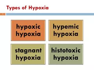
Classification Etiology Pathogenesis
发生缺氧的基本环节 Atmosphere Decreased PiO2 Ventilation & Diffusion Hypotonic hypoxia External respiratory dysfunction Binding with Haemoglobin Transportation of gases in blood Venous-to-arterial shunts Taking and utilization of oxygen in tissue cells
Hypotonic hypoxia is characterized by the decrease of PaO2 (<60mmHg), also called hypoxic hypoxia. Etiology • Decreased PO2of inspired air: Atmospheric hypoxia(大气性缺氧) Plateau (高原性缺氧) Hypotonic hypoxia(低张性缺氧)
External respiratory dysfunction: respiratory hypoxia (呼吸性缺氧) • Venous-to-arterial shunts:tetralogy of Fallot 法洛氏四联症(tetralogy of Fallot )
Characteristics of blood O2 ↓ ↓ N PaO2 SO2 CO2maxCaO2 A-VdO2 ↓/ N ↓ Cyanosis (紫绀):Cyanosis is a bluish or purplish tinge to the skin and mucous membranes. Approximately over 5g/dL of unoxygenated hemoglobin in the capillaries generates the dark blue color appreciated clinically as cyanosis. O2 diffusion in tissue
Atmosphere Decreased PiO2 Ventilation & Diffusion External respiratory dysfunction Hypotonic hypoxia Binding with Haemoglobin Transportation of gases in blood Venous-to-arterial shunts Taking and utilization of oxygen in tissue cells Hemic hypoxia Abnormality of HB
Hemic hypoxia 血液性缺氧 Hemic hypoxia refers to the altered affinity of HB for oxygen or decrease in amount of HB in the blood, also can be termed as isotonic hypoxemia (等张性低氧血症)。 Etiology • Anemia:贫血性缺氧(anemic hypoxia) • Carbon monoxide poisoning:碳氧血红蛋白(HbCO) • Methemoglobinemia:HbFe3+OH;肠源性紫绀
Carbon monoxide poisoning CO combines preferentially with hemoglobin to produce COHb, displacing oxygen and reducing systemic arterial oxygen (O2) content. CO binds reversibly to hemoglobin with an affinity 200- 230 times that of oxygen. Consequently, relatively minute concentrations of the gas in the environment can result in toxic concentrations in human blood. Possible mechanisms of toxicity include: • Decrease in the oxygen carrying capacity of blood. • Alteration of the dissociation characteristics of oxyhemoglobin, further decreasing oxygen delivery to the tissues. • Decrease in cellular respiration by binding with cytochrome a3. • Binding to myoglobin, potentially causing myocardial and skeletal muscle dysfunction.
(2)一氧化碳中毒(Carbon monoxide poisoning) 吸入 CO CO + Hb 碳氧血红蛋白(HbCO) 2,3-DPG生成↓ 氧离曲线左移 Hb失去携O2能力 HbO2释放氧↓ 组织缺氧 CO + 1个Hb亚单位 Hb产生变构 其余3个血红素结合的氧也不易释放 氧离曲线左移 Four binding sites ※CO与Hb的亲和力比O2大210倍,当吸入的气体内含有0.1%CO时,血液中的血红蛋白可能有50%转为HbCO。
Methemoglobinemia, MHb • Definition: Methemoglobinemia is a disorder characterized by the presence of a higher than normal level of methemoglobin (metHb) in the blood,in which the ferrous (2+) form of heme is oxidized to the ferric form (3+) thus making the heme moiety unable to bind oxygen. In addition, the remaining monomers of ferrous heme within a hemoglobin tetramer bind their oxygen more tightly causing a left shift of the oxygen dissociation curve and reduced oxygen delivery at the tissue level.
Causes • Hereditary/Congenital:Hemoglobin M Cytochrome b5 reductase deficiency (NADHdeficiency)—responsible for 95% of MetHgb reduction, NADPH deficiency of the HMP shunt Acquired: Multiple drugs and toxins including aniline dyes, benzene, chloroquine, dapsone, local anesthetic agents, reglan, naphthalene, nitrites (including NTG and NO), primaquine, phenazopyridine, and sulfonamides.
Clinical presentation: • Chronic methemoglobinemia: chronically elevated levels of MetHgb often are asymptomatic or present with headache, fatiguability, or “slate blue skin” complaints. • Acquired (acute) methemoglobinemia: typically symptomatic due to lack of compensatory mechanisms: cyanosis, dyspnea, fatigue, lethargy, AMS, shock, seizures and death. Severity depends on percent methemoglobinemia. (1% is normal) 3-15% skin discoloration 20% cyanosis or asx 25-50%, HA, lightheaded, weak, chest pain, confusion 50-70% delirium, seizure, lactic acidosis >70% arrhythmia and death.
Characteristics of blood O2 PaO2 SO2 CO2maxCaO2 A-VdO2 ↓ N /N N ↓ /N ↓ Color of skin • Anemia:Pale skin color • HbCO:Classic cherry red skin is rare (ie, "When you're cherry red, you're dead"); pallor is present more often. • HbFe3+OH:Bluish coloring, cyanosis 贫血的氧离曲线
70% 70% 20% normal methemoglobin concentration Note the chocolate brown color of methemoglobinemia. This dark hue imparts clinical cyanosis when methemoglobin levels are at 1.5 g/dL (approximately 10-15% methemoglobin concentration); however, a level of 5 g/dL of deoxygenated blood is required for similar effects. Therefore, when methemoglobin levels are relatively low, cyanosis may be observed without cardiopulmonary symptoms.
Atmosphere Decreased PiO2 Ventilation & Diffusion External respiratory dysfunction Hypotonic hypoxia Binding with Haemoglobin Abnormality of HB Hemic hypoxia Transportation of gases in blood Venous-to-arterial shunts Taking and utilization of oxygen in tissue cells Circulatory hypoxia hypokinetic
Circulatory hypoxia循环性缺氧 Circulatory hypoxia refers to inadequate blood flow leads to inadequate oxygenation of the tissues. 1.Tissue ischemia:shock, heart failure 2.Tissue congestion:venous embolism Etiology • Systemic:shock, heart failure • Local:embolism, atherosclerosis
Characteristics of blood O2 PaO2 SO2 CO2maxCaO2 A-VdO2 N N N N ↑ Color of skin • Tissue ischemia:pale • Tissue congestion:cyanosis
Atmosphere Decreased PiO2 Ventilation & Diffusion External respiratory dysfunction Hypotonic hypoxia Binding with Haemoglobin Abnormality of HB Hemic hypoxia Transportation of gases in blood Venous-to-arterial shunts hypokinetic Circulatory hypoxia Taking and utilization of oxygen in tissue cells Histogenous hypoxia Dysfunction of O2 utilization
Histogenous hypoxia组织性缺氧 Histogenous hypoxia refers to the tissue cell cannot make use of the O2 supplied to them. Etiology • Tissue poisoning:histotoxic hypoxia cyanide, arsenide, sulphide • Vitamin insufficiency:vitaminB1, B2, PP • Mitochondrial damage:radiation, bacteria、uremia
Characteristics of blood O2 PaO2 SO2 CO2maxCaO2 A-VdO2 N N N ↓ N Color of skin • Deceptively healthy pink to red skin color
急性严重缺氧时机体变化以失代偿和损伤为主;急性严重缺氧时机体变化以失代偿和损伤为主; 轻度缺氧时机体或细胞以代偿性调节为主。 慢性缺氧时机体的代偿反应和缺氧性损伤并存。
Compensatory reaction 代偿性反应
PaO2<60mmHg(8kPa)刺激外周化学感受器 呼吸中枢(+) 呼吸加深加快 窦神经、迷走神经 PaO2↑ PAO2和PaO2↑ C.O和肺血流量↑ 静脉回流↑ 胸内负压↑ 组织供氧↑ 有利于氧的摄取和运输 PaCO2 ↑ 刺激中枢化学感受器 Respiratory system 急性缺氧时最主要的代偿反应--呼吸功能增强使肺通气量增加
呼吸性碱中毒 中枢化学感受器兴奋性↓ 【早期】 高原地区 呼吸加深加快 肺通气量增加65% 【数日后】 高原地区 呼吸加深加快 肺通气量增加5~7倍 数日后 呼碱被纠正 中枢化学感受器(+)↑ 【长期居住】 肺通气量接近正常(肺通气量增加15%) 外周化学感受器对缺氧的敏感性↓ 机体对缺氧的耐受性↑ 肺通气变化与缺氧持续时间的关系
(1)心输出量增加 交感N(+) 心率↑;心肌收缩性↑;静脉回流↑ C.O ↑ (2)肺血管收缩(Kv通道为主) 缺氧 Kv通道开放↓K+外流↓ 膜电位↑ 平滑肌(+)↑ Ca2+内流↑ 肺内合成释放缩血管物质大于扩血管物质 肺小动脉收缩 交感神经兴奋↑ Circulatory system (3)血液重分布 脑、心血管平滑肌以KCa、KATP通道为主。 (4)毛细血管增生
Hematologic system 1)红细胞增多 1)红细胞增多 2)氧合血红蛋白解离曲线右移 • 急性缺氧 脾脏、肝脏收缩,将储存血液释放入体循环,增加氧的摄取和运输能力 • 慢性缺氧 血浆中ESF(EPO)增加,使红细胞数和Hb量明显增加 • 可增加血液的氧容量和氧含量 • 过度增加可使血粘度增加 2)氧合血红蛋白解离曲线右移 缺氧时红细胞内2,3-DPG增多,2,3-DPG增多使氧离曲线右移 • 有利于HbO2在组织部位释放出较多的氧 • 当paO2低于8kPa时,氧离曲线右移会明显影响肺部血液对氧的摄取 2,3-DPG的生成与分解
Tissues and cells • 细胞内呼吸功能增强:线粒体数目,呼吸酶活性 • 糖酵解增强 • 肌红蛋白增加:与氧的亲和力增加 • 低代谢状态
Hypoxia inducible factor-1(HIF-1) VEGF, EPO, glycolysis enzymes NATURE REVIEWS | Immunology volume 9 | September 2009
Hypoxia Injury 缺氧对机体的损伤性作用
Hypoxia / ischemia Mitochondrion ↓Oxidative phosphorylation ↓ATP ↓Na pump ↑Anaerobic glycolysis Other effects Detachment of ribosome, etc. ↑Influx of Ca2+, H2O, and Na+ ↑Efflux of K+ ↓Glycogen ↓pH Clumping of nuclear chromatin ↓Protein synthesis ER swelling Cellular swelling Loss of microvilli Blebs Lipid deposition