Innovative Imaging Solutions for Biomedical Data Analysis
140 likes | 166 Vues
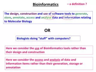
Explore cutting-edge techniques in image acquisition and processing for non-invasive detection of medical conditions such as coronary artery anomalies and liver cancer. Utilizing novel wavelet technology and isotropic wavelet-based texture detection, our research aims to revolutionize medical imaging for early diagnosis and improved patient care.

Innovative Imaging Solutions for Biomedical Data Analysis
E N D
Presentation Transcript
Manos Papadakis, with Bernhard G. Bodmann, Donald J. Kouri, Department of Mathematics Institute for Digital Informatics and Analysis, Texas Learning and Computation Center University of Houston Mathematics-Bioinformatics
Collaboration Members Image Analysis: B. G. Bodmann, PhD; M. Papadakis, PhD; D. K. Hoffman, PhD; D. J. Kouri, PhD; Bingxin Li, Indika Walimuni, Amrendra Vijay, PhD; G. Gogoshin, Alex Gittens; N. Karayiannis THI Collaborators: S. W. Casscells, MD; S. D. Gertz, MD; W. T. Wilner, PhD; J. L. Conyers, PhD; I. Aboshady, MD; D. Vela, MD; P. Cherukuri (now at Rice University); S. Lukovenkov, MD; R. M. Mazraeshadi, MD MDACC Collaborators: D. Cody, PhD; G. Gladish, MD;E. Johnson Baylor CM: Claudia Robertson, MD, Ph.D, Alex Valadka, MD
Overview Biomedical Data Processing and Handling: Image Acquisition, and post-processing Storage and Exchange of Information Creation of Data Bases Automated Assisting Diagnostic Tools Our group focuses on: Post-Processing Improving the Image Acquisition Targeted applications: Non-invasive detection of anomalies in coronary arteries Non-invasive early detection of liver cancer Non-invasive estimation of Cerebral Blood Flow with portable X-ray CT-scanners
Imaging problem: Calcium scoring is only a predictor of AMI. The primary factor is to detect vulnerable plaque, plaque which is likely to rapture. This tissue type is soft and lightly calcified and causes Acute Myocardial Infarction, which kills without warning. Solution: Identify tissue which is anomalous, different from the surrounding normal. Targeting: Coronary Arteries and carotid arteries Our techniques apply to all modalities (X-ray, MRI, IVUS) We currently work with X-ray CT scans.
Novel Wavelets, Without Directional Preference old, w/directionalpreference newno preferred direction Plots of intensity values for old and new two-dimensional wavelets. First Generation Isotropic Wavelets
Why Isotropic Wavelets Should be on a Radiologist’s Wish List • Both work in 3D (+time, if desired) with Fast Algorithms • Have no preferred directions just as human vision. • Can distinguish a variety of features in images. Use them to produce “digital contrast agent” • Establish tissue characterization by isotropic wavelet-based texture detection.
Anomalous content Original data (vox: 27mu ) Apply statistics to the of wavelet-encoded Image to discriminate different types of tissue 3 1. Select reference tissue for wavelet adaptation 2. Remove reference- type tissue 2. Remove reference- type tissue Data obtained from coronaryspecimen withMicro CT Scanner.
Wavelet-based tissue segmentation, lipid calcified
HU intensities vs. 3D-Isotropic Wavelets Original data viewed in slices Processed data superimposed 3D rendering threshold-based wavelet-based
Recent improvements in resolution – great! • Semi-automated post-acquisitional processing that “stains” lesions. • Isotropic Wavelets. Wish List vs. Recent Progress • CT images as good and clean as histology • Automated detection and indication of diagnostically relevant tissue types. • Use of clinical CT-scanners (X-ray,MRI) • Fast processing, no directional preference
Current Research Goals Validate the New Digital Contrast Agent using: 64-slice CT-scanners and Flat-Panel CT Investigate potential application in liver cancer detection (Data supplied by GE) Assess performance with Flat Panel and with reduced radiation.
Benefits • Non-invasive monitoring of patients who have stents. • Non-invasive screening of general population to assess • of acute myocardial infarction • Earlydetection of cancerous formations
Grant Acknowledgement NSF Applied Mathematics Program T5-UT Health Science Center @Houston Total-Elf-Fina (why not do the same tricks to find oil !!!) TLCC-UH