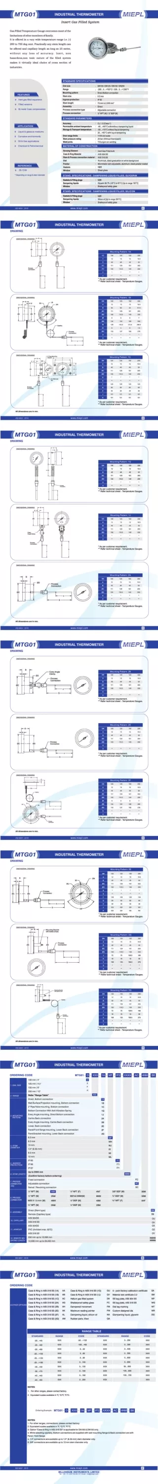
Download
1 / 28
280 likes | 456 Vues
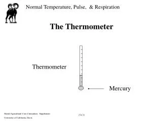
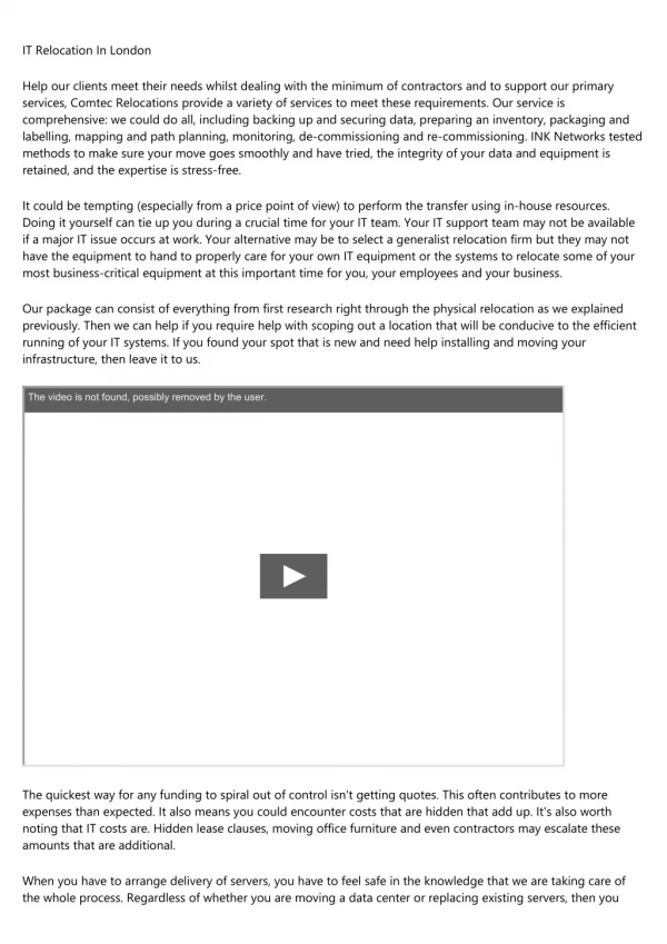
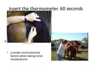
Insert the thermometer. 60 seconds . consider environmental factors when taking rectal temperatures. A, The thermometer has been inserted and secured with the clip to the tail hairs. B, Thermometer secured to hair coat with the clip. . Pulse Rate/Heart Rate.

E N D
Insert the thermometer. 60 seconds consider environmental factors when taking rectal temperatures
A, The thermometer has been inserted and secured with the clip to the tail hairs. • B, Thermometer secured to hair coat with the clip.
Pulse Rate/Heart Rate • The pulse rate is taken by palpation of arteries • Heart rate and pulse rate are nor the same: heart rate refers to the number of heart bests/minute (bpm); pulse rate refers to the number of palpable arterial pulse waves/minute • In normal animals heart rate and pulse rate are equal • Arterial pulses may be palpated at several locations • Pulse deficit (heart rate ↑ pulse rate↓) • Pulse is described as strong, bounding, weak, thready, or other non-specific terms
Facial artery is the most convenient location where it courses over the ventral aspect of the mandible, rostral to the origin of the masseter muscle
B, Identify the facial artery along the medial aspect of the mandible. • C, Press the vascular bundle against the medial aspect of the mandible
Transverse facial artery • Is located in a horizontal depression about 1 inch caudal to the lateral canthus of the eye and just below the zygomatic arch.
Coccygeal artery • Supplies the tail and is located along the ventral midline of the tail.
Dorsal metatarsal artery • Is located between the metatarsal 3 and 4 (cannon bone and lateral splint bone) on the hind limp
E, Location of the lateral digital artery over the lateral proximal sesamoid bone and proximal to the lateral collateral cartilage. F, Palpation of the digital arteries over the proximal sesamoid bones. G, Palpation of the digital arteries proximal to the collateral cartilages.
Reparatory rate The number of respirations/minute can be counted in several ways: • Using a stethoscope to listen to air movement in the trachea or chest • Using a hand to feel movement of air in and out of a nostril • Simply counting chest excursions (rise and fall of the thoracic wall)/minute
Heart Auscultation • Horses are athletes: the heart of the average horse may be as large as a basketball. • Auscultation may be done on the left or right side of the chest, though most of the heart valves and sounds are heard best from the left side • The most common cause of an irregular heart rhythm in the horse is the second degree atrioventricular (A-V) block
Landmarks for the heart • The horizontal marks indicate the level of the shoulder and elbow joints. The vertical mark indicates the caudal border of the triceps muscle.
Auscultating the heart. • A, Gently lift the triceps muscle away from the chest wall. • B, Place stethoscope against the chest wall, deep to the triceps muscle.
Video http://www.youtube.com/watch?v=Y6CyG-cfyzI&feature=related
Landmarks for the lung.Borders of the left lung field for auscultation
Lung auscultation The stethoscope is placed in several locations within the lung field to listen to several breaths at each location
Video • http://www.merckvetmanual.com/mvm/htm/bc/reshs921.htm
Abdominal Auscultation • A stethoscope is used to listen to abdominal sounds, which are created by movements of the intestines • This commonly referred to as gastrointestinal motility or GI motility Landmarks for abdominal auscultation in the flank area are the point of the hip (tuber coxae) and the last rib.
Abdominal Auscultation(cont’d) Should listen in each quadrant (4) for at least 1 minute each, on left and right side 0 = no motility +1 = hypomotility +2 = normal motility +3 = hypermotility
Standard four point auscultation A, Auscultation of the upper left abdominal quadrant. B, Auscultation of the lower left abdominal quadrant. C, Auscultation of the upper right upper abdominal quadrant. D, Auscultation of the lower right abdominal quadrant.
Mucous membranes • Mucous membranes are tissues that have the ability to make and secrete mucus. • Mucus membranes’ color is helpful for disease diagnosis • Cyanosis is bluish coloration- low oxygen of the blood • Brick red coloration indicates septicemia or shock or both, colic, endotoxemia • Purple gum line indicates endotoxic shock • Yellowish coloration of the gum indicates icterus • Pale mucus indicates anemia
Examination of mucous membranes • .A, Examination of the gums. • B, Examination of the conjunctiva
Examination of mucous membranes • C, Examination of the mucosa of the nares. • D, Examination of the vulva in the female.
Hydration Status Skin turgor test At the point of the shoulder 1 second or less is normal 1 second or more- >5% >8 seconds: Severely dehydrated Capillary refill time Less than 2 seconds Dehydration; shock 5 to 8 seconds
Neurological Examination Confirm disease Find where More tests usually needed