Download
1 / 57
580 likes | 836 Vues
Diagnostic Nuclear Physics in Medicine. Thomas J. Ruth UBC/TRIUMF PET Program TRIUMF, Fall 2004. Diagnostic medicine tries to look into the body to see what is happening, in life and in death. Rembrandt - anatomy lesion. How can we probe the human body without a knife?

E N D
Diagnostic Nuclear Physics in Medicine Thomas J. Ruth UBC/TRIUMF PET Program TRIUMF, Fall 2004
Diagnostic medicine tries to look into the body to see what is happening, in life and in death. Rembrandt - anatomy lesion
How can we probe the human body without a knife? But first some fundamental ideas that are needed to understand this topic.
Size matters! How big is Avogadro’s number – 6.02 x 10²³? Each Smartie – 1 cm3 or 1 mL 1 mole of Smarties = 6 x 10²³ mL Assume a truck with a capacity of 90 m3 (10x3x3) = 9 x 10⁷ mL The number of trucks needed – 6 x 10¹⁵ If each truck is 13 m long, that means that lined up they would be 7.8 x 10¹³ km in length or 200 million earth to moon distances.
1 grain of salt – 0.12 mg = 2 x 10⁻⁶ moles (or 2 micromoles) = 3 x 10¹⁷ molecules This issue will come up later in our discussion of tracers.
Normal chest x-ray X-ray images produce shadows based on density of matter.
All of the images shown thus far look at structure. Many of these images would look the same in a living person or in a cadaver. What about function?
Why Use PET Imaging? • PET imaging is capable of providing quantitative information about biochemical and physiological processes, in vivo.
Tracer Principle • Tracer behaves in a similar way to the components of the system to be probed. • Tracer does not alter the system in any measurable fashion. • Tracer concentration can be measured.
Basic Principles of PET • Based on tracer principle. • Tracer labeled with positron emitting radioisotope. • Positron decay. • Coincidence detection of annihilation radiation.
Positron Annihilation Positron emitter Positron range 1 - 10 mm Neighboring atom g #2 g #1 Gamma ray range 10 mm - 180°
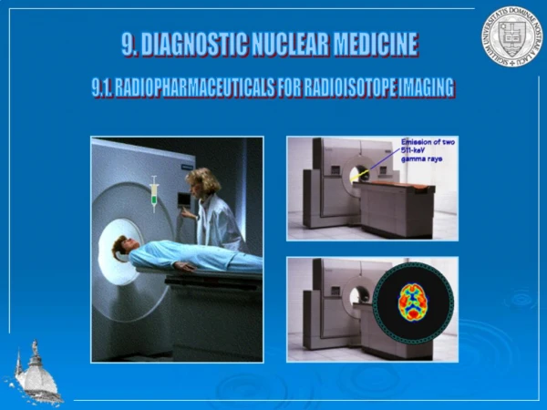
Positron Emission Tomography detector #2 g #2 g #1 detector#1
PET measures concentration of radioactivity 1) Gamma rays escape from the body: Allows external detection. 2) Two gamma rays are emitted at 180° when a positron annihilates: The annihilation occurred somewhere along the line of response (LOR) between the 2 detectors.
PET measures concentration of radioactivity 3)Regions with greater radioactivity levels produce more LORs: Concentration of radioactivity can be measured and quantified.
Corrections necessary for quantitative PET (mCi/cc tissue) • Scatter correction • Attenuation correction • Random subtraction • Normalization • Calibration
CH2OH CH2OH O O OH OH OH OH OH OH OH F* Fluorodeoxyglucose (FDG) Glucose
FDG Uptake and Retention Cells Blood Glycolysis Glucose Glucose Glucose-6P FDG FDG FDG-6P
The brain, my second most favorite organ. Woody Allen
Goals of the Pacific Parkinson’s Research Centre • Determine the origins of PD • Follow natural history of disease (Progression) • Develop treatments • Control complications of treatment
Clusters • CBC, Vancouver – 4/120 • Community college, eastern BC – 4/32 • Garment factory, Montreal – 3/8 200-300 per 100,000 population. ~ 1% in population aged over 60 years.
Radiopharmaceuticals • Dopamine system • 18F-FDOPA • 11C-Methylphenidate • 11C-Dihydrotetrabenazine • 11C-Raclopride • 11C-Sch23390 NB – 11C high specific activities (10 mCi @ 10 Ci/mmole ~ 1014 molecules)
For PET research you need clever and gifted chemists! Photo by Will Brown/courtesy of Chemical Heritage Foundation
Controls p=0.0274 MPTP-exposed Exposure 1st scan 2nd scan
11C-DTBZ 11C-SCH 23390 11C-MP D1R VMAT2 Tyr DOPA DA D2/D3R DAT 11C-RACLOPRIDE 18F-FD 18F-FD 11C-MP 11C-DTBZ
DTBZ vs FDOPA DTBZ vs MP 11C-DTBZ 11C-SCH 23390 (n = 35) (n = 35) 100 100 11C-MP D1R 75 75 VMAT2 MP (% Normal) 50 50 FDOPA (% Normal) Tyr DOPA DA D2/D3R 25 25 DAT 11C-RACLOPRIDE 0 0 0 25 50 75 100 0 25 50 75 100 18F-FD DTBZ (% Normal) DTBZ (% Normal) Investigation of disease compensatory mechanisms Lee et al 2000
The importance of isomeric purity. VMAT2 - 11C-dihydrotetrabenazine Multiple chiral centres
In vivo assessment of endogenous DA concentration DA DA DA 11C-Raclopride Endogenous DA competes with raclopride for the D2 receptor
Apomorphine-induced changes in RAC binding (putamen) 3 Error Bars: ± 1 Standard Error(s) 2.5 2 1.5 1 .5 0 APO_0 APO_1 APO_2
Can you focus this please? Circa 1976, BNL/UPenn
Yes! • 25 years later • And 100’s M $
Crystal material: LSO/LYSO Crystal size 2.1 x 2.1 x 10 mm3
High Resolution Research Tomograph • 119,000 detector elements • 4,000,000,000 lines of response • > 1Gbyte of data per image frame
Significant Results Beyond UBC/TRIUMF PET FDG Supply (b-HC!) 18F- FDG mPET PET/CT Vendor not selected!