Scapular, Parascapular and P ectoralis Flaps
390 likes | 2.49k Vues
Scapular, Parascapular and P ectoralis Flaps. Ian Maxwell Summer anatomy July 4, 2013. Mathes and Nahai muscle flap classification. Mathes and Nahai classification of fasciocutaneous flaps: . Cormack and Lamberty classification of fasciocutaneous flaps:.

Scapular, Parascapular and P ectoralis Flaps
E N D
Presentation Transcript
Scapular, Parascapularand Pectoralis Flaps Ian Maxwell Summer anatomy July 4, 2013
Cormack and Lamberty classification of fasciocutaneous flaps:
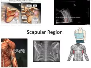
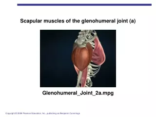
Triangular and Quadrangular spaces: • Triangular space: • Borders are: • Teres minor/subscap • Teres major • Long head triceps -CIRCUMFLEX SCAPULAR ARTERY • Triangular interval • Borders are: • Teres major • Long head triceps • Lat head tripceps/humerus -PROFUNDA BRACHII ARTERY - RADIAL NERVE • Quadrangular space • Borders are: • Teres minor/subscap • Long head triceps • Lat head triceps/ humerus • Teres major -POSTERIOR HUMERAL CIRCUMFLEX ARTERY -AXILLARY NERVE
Classification: MathesandNahai=TypeBseptocutaneous CormackandLamberty=TypeBsinglefasciocutaneousperforator Arterial Supply: ScapularFlap=Transversebranch ofCircumflexScapularartery ParascapularFlap=Descendingbranch ofCircumflexScapularartery VenousDrainage=venaecomitantes NOTE:SubscapularVeinnotpaired Nerve: Noreliable cutaneousnerve
SkinPaddle • Scapular=10cmx25cm • Parascapular=15cmx25cm LargerifSTSGdonor…notideal PedicleLength/Diameter • • • • Transversebranch=4-9cm/1.5-2.0mm Descendingbranch=4-6cm/1.5-2.0mm CircumflexScapular=7-10cm/2.5-3.5mm Subscapular=11-14cm/3.5-4.5mm NOTE:cancombine scapular and parascapularpaddlesifboth vesselsincluded
BoneFlap • 2cmwidex10cmlong • Atleast2cmfromglenoid • Includemuscletopreserve periostealbloodsupply Tipissuppliedby angular branchfromthoracadorsal artery
FlapApplications LocalPedicledFlap • Shoulder • Axilla FreeFlap • • • Head,neck,oralcavity/mandible Upperextremityandhand Lowerextremityandfoot Chimericflap • • • • Skin Fascia Muscle Bone
FlapElevation ScapulaFlap Incisefasciato protectpedicle Markflapto include triangularspace Dissectmedial tolateral, suprafascial
FlapElevation ScapulaFlap Foradditional pediclelengthand vesselcalibermust dissectthrough triangularspaceto subscapularartery
FlapElevation ParascapulraFlap Incisefasciato protectpedicle Markflapto include triangularspace Dissectinferior tosuperior suprafascial
FlapElevation ParascapulaFlap Foradditional pediclelengthand vesselcalibermust dissectthrough triangularspaceto subscapularartery
Classification • Mathes and Nahai type V (segmental and dominant) • Variations: • Muscle flap (most common) • Musculocutaneous • Osteomusculocutaneous (rib or sternum) • chondromusculocutaneous
Applications • Face, Oral cavity, Neck, • Sternum/mediastinum, • Axilla and shoulder • Upper extremity functional muscle transfer • Reconstruction of • mandible, esophagus, breast, functional muscle for elbow
Pectoralis Major Muscle anatomy • Origin: • Sternal head: first 7 ribs and sternum and aponeurosis of external oblique • Clavicular head: medial head of clavicle • Insertion: • Lateral lip of bicipital groove of humerus
Arterial supply of Pec Major flap: • Major pedicle • Pectoral branch of thoracoacromial artery • Length = 4.5cm • Diameter = 2-3mm • Minor/segmental pedicles • Medially: Intercostal perforators 1-6 • Usually pedicled off first 2 (deltopec flap) • Length = 1-2cm • Diameter <1mm • Laterally: pec branch of lateral thoracic artery • Length = 3cm • Diameter = 1-2mm • Venous drainage via venae comittantes
Innervation • Medial pectoral nerve (motor) • Lateral pectoral nerve (motor) • Intercostal nerves 2-7
Pec branch of Thoracoacromial artery Sternocostal head Lateral thoracic artery Clavicular head Clavicular Branch of thoracoacromial a.
Flap harvest: • Incision: • Midline sternotomy: for sternum reconstruction • Subclavicular: usually for head and neck coverage • Through skin island if one is planned • Dissection: through pre-pectoral plane raise skin and subcu tissue off of pec
Flap Harvest • Turnover (for sternum) • Divide through pec laterally • Dissect under pec lateral to medial until sufficient turnover possible based on IMA perforators • Skin paddle • Axis of rotation is line from acromion to xiphoid • Skin paddle of 8x10 cm usual limit for 1o closure, design over pec muscle, +/- dopplering of perforators
Skin paddle cont. • Dissection proceeds as for turnover: • Divide lateral pec and insertion at ext oblique (including lateral pec artery) • Divide medial sternocostal origin • Dissect pec major away from pec minor and chest wall inferior to superior • Pedicle lies medial to pec minor on underside of pec major • Flap is tunneled to desired location
Functional muscle transfer • Incision along anterior axillary line and dissect skin off muscle • An innervated portion of muscle outlined • Divide origin at sternum and clavicle • Preserve thoracoacromial pedicle and motor nerves • Tunnel muscle through axilla and suture to biceps tendon
Deltopectoral flap • Fasciocutaneous flap based on 2nd or 3rd perforating branches of IMA • More commonly used for head and neck recon given medial pedicle
References • Microsurgeon.org • Serafin, d. Atlas of microsurgical tissue transplantation • Wei, Mardini. Flaps and reconstructive surgery • Halifax flaps manual • Ash’s scapular/parascapular talk last year