Download
1 / 1
10 likes | 81 Vues
Microscopic analysis of Noncystic PWMI in white matter junction of parietal and occipital lobes in preterm and very preterm cases. Hemalun-phloxine staining, astrocytes, microglia, and axons highlighted.

E N D
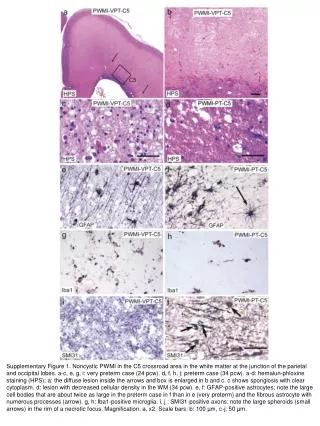
Supplementary Figure 1. Noncystic PWMI in the C5 crossroad area in the white matter at the junction of the parietal and occipital lobes. a-c, e, g, i: very preterm case (24 pcw). d, f, h, j: preterm case (34 pcw). a-d: hemalun-phloxine staining (HPS); a: the diffuse lesion inside the arrows and box is enlarged in b and c. c shows spongiosis with clear cytoplasm. d: lesion with decreased cellular density in the WM (34 pcw). e, f: GFAP-positive astrocytes; note the large cell bodies that are about twice as large in the preterm case in f than in e (very preterm) and the fibrous astrocyte with numerous processes (arrow). g, h: Iba1-positive microglia. i, j : SMI31-positive axons: note the large spheroids (small arrows) in the rim of a necrotic focus. Magnification: a, x2. Scale bars: b: 100 µm, c-j: 50 µm.