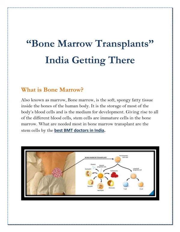
Download
1 / 45
450 likes | 606 Vues
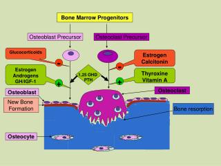
Renal pathology in other solid organ and bone marrow transplants. Anke Schwarz, Deptm. of Nephrology Verena Bröcker, Inst. for Pathology. NHL 1997 BMT 1997 Lung Tx 2001 ESRD 2011 37 yrs old. Changes in reciprocal s-creatinine ( ~GFR) in cardiac-transplant recipients.

E N D
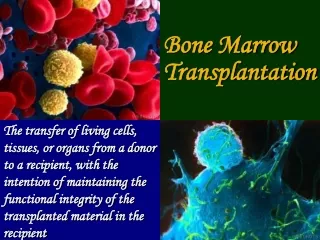
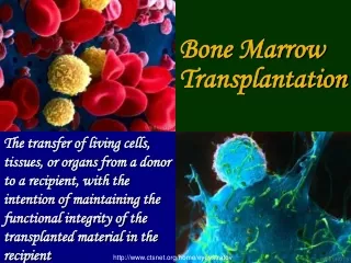
Renal pathology in other solid organ and bone marrow transplants Anke Schwarz, Deptm. of Nephrology Verena Bröcker, Inst. for Pathology
NHL 1997 BMT 1997 Lung Tx 2001 ESRD 2011 37 yrs old
Changes in reciprocal s-creatinine (~GFR)in cardiac-transplant recipients Myers et al, NEJM 1984
Evolution of GFR in heart-transplanted patients (n=151) Lindelöw et al, JASN 2000
Seriel creatinine clearances in patients after lung or lung-heart transplantation (n=115) Filled boxes=later ESRD Kunst et al, J Heart Lung Transplant 2004
Survival at dialysis after lung transplantationcompared with expected survival (n=40) Mason et al, J Heart Lung Transplant 2007
Renal biopsies after non-renal transplantation • 105 renal biopsies in 101 patients 2000 – 2009 (Hannover) • 14 after bone marrow transplantation • 41 after liver transplantation • 30 after lung transplantation • 20 after heart transplantation
Renal biopsies after non-renal transplantation • 105 renal biopsies in 101 patients 2000 – 2009 (Hannover) • 14 after bone marrow transplantation • 41 after liver transplantation • 30 after lung transplantation • 20 after heart transplantation • 34 renal biopsies in 34 patients 1987 – 2000 (Basel) • 20 after bone marrow transplantation • 14 after heart transplantation
Renal biopsies after non-renal transplantation • Indication for renal biopsy in Hannover: • non-recovery after ARF at transplant. 9% • creatinine increase 83% • rapid creatinine increase 22% • proteinuria 22% • nephrotic syndrome 13% • evaluation of renal prognosis before • re-transplantation of an extrarenal organ 9%
Renal biopsies after non-renal transplantation • End stage renal disease in Hannover: • BM: 1 out of 1375 patients (0.07%) • Liver: 32 out of 2016 patients (1.6%) • Lung: 35 out of 725 patients (4.8%) • Heart: 41 out of 505 patients (8.1%)
101 patients with renal biopsies after non-renal transplantation
101 patients with renal biopsies after non-renal transplantation
34 renal biopsies after heart transplantation P=0.01 NS NS
Main histopathological lesions in kidney biopsies * (n=101) * Interlobular/ arcuata-arteries
Glomerulosclerosis and fibrosis in organ groups (GS) (IF/TA)
Chronic vascular lesions in organ groups (AH) (AS) p=0.006
Glomerulonephritis and TMA in organ groups Bone marrow: SLE, MGN, fibrillary GN Liver: IgA, MPGN Heart: IgA, Shunt-Nephritis
Interstitial fibrosis/ tubular atrophy and kidney outcome >/= 3 years follow up
Main histopathological diagnosis (n=101)
Main histopathological diagnosis • “NOS” • No reason for impaired renal function • IFTA>20% and GS and nephrosclerosis without obvious reason (n=101) • “Other” • Bone marrow: Nephrocalcinosis, Minimal Change, Cast-Nephropathy • Heart: Nephrocalcinosis/ interstitial Nephritis • Liver: Diabetic NP, Iron-overload, Minimal Change • Lung: Polyoma-Virus NP, Nephrocalcinosis
Kidney biopsies of heart transplanted patients Hannover and Basel
Kidney biopsies of heart transplanted patients Hannover and Basel
Thrombotic microangiopathy • Hannover 2000 – 2009 n=10/101 (10%) • TMA after BM-Tx: n=1 • TMA after liver-Tx: n=5 • TMA after lung-Tx: n=4 • TMA after heart-Tx: n=0 • Basel 1987 – 2000 n=8/34 (24%) • TMA after BM-Tx: n=5 • TMA after heart-Tx: n=3
TMA • TMA in 428 renal transplant patients 1.2% • TMA in 4203 native kidneys 1.1% • TMA in 101 native kidneys after non-renal transplantation in Hannover 2000-2009 10% • TMA in 14 native kidneys after non-renal transplantation in Basel 1987-2000 24%
TMA after non-renal transplantation • Shulman et al 1981 BM 3 autopsy cases • Bonser et al 1984 liver case report • Dische et al 1988 liver case report • Nizze et al 1988 BM, heart 167 autopsies, 15% • Griffiths et al 1996 heart, lung 22 histologies, 14% • Pillebout et al 2005 liver 15 histologies, 50% • Lefaucheur et al 2008 liver 26 histologies, 47%
Thrombotic microangiopathy • Complement mutation-associated • de novo Thrombotic Microangiopathy • following kidney transplantation M. Le Quintrec et al AJT 2008 TMA with HUS after RTX n=24 Mutations in genes encoding complement abnormalities 29%
Thrombotic microangiopathy • Immunosuppression with TMA in Hannover: • cyclosporine-based n=5 • tacrolimus-based n=2 • cyclosporine and everolimus n=3
arteriolar lesion 3 glomerular lesion 5 (glomerular & arteriolar lesion 3)
Thrombotic microangiopathy • Symptoms of TMA in Hannover n=10: • hemolytic uremic syndrome 5 • severe hypertensive episode 6 • lung edema 1 • retinal bleeding 1 • creatinine increase 8 • (proteinuria 4g/day 1)
6 month kidney survival after biopsy TMA p=0.01
Renal biopsies after non-renal transplantation • most often signs of acute renal failure, pre-existing hypertension and CNI-toxicity • high number of patients with glomerulonephritis • high number of patients with TMA Conclusion 1
Renal biopsies after non-renal transplantation • mostly signs of acute renal failure, pre-existing hypertension and CNI-toxicity • high number of patients with glomerulonephritis • high number of patients with TMA Conclusion 1
Renal biopsies after non-renal transplantation • mostly signs of acute renal failure, pre-existing hypertension and CNI-toxicity • high number of patients with glomerulonephritis • high number of patients with TMA Conclusion 1
Renal biopsies after non-renal transplantation • find early specific lesions • in the advanced stage the extent of IFTA determines renal outcome • extrarenal transplantation may accelerate the process of „aging“ by multifactorial causes Conclusion 2
Renal biopsies after non-renal transplantation • find early specific lesions • in the advanced stage the extent of IFTA determines renal outcome • extrarenal transplantation may accelerate the process of „aging“ by multifactorial causes Conclusion 2
Renal biopsies after non-renal transplantation • find early specific lesions • in the advanced stage the extent of IFTA determines renal outcome • extrarenal transplantation may accelerate the process of „aging“ by multifactorial causes Conclusion 2
Therapeutic consequences • Reduce CNI as much as possible • Finish CNI - mTOR inhibitor combination • Agressive blood pressure control • Plasmapheresis in TMA with HUS • Renal replacement planning
Renal transplantation by LIVING DONATION
Thank you for your attention • Department of Nephrology • Hermann Haller • Anke Schwarz • Institute for Pathology • Hans Kreipe • Jan U. Becker • Verena Bröcker • Hemato-Oncology • Gastro-Enterology • Pulmonology • Thoracic Surgery • Christian Koenecke • Jens Gottlieb • Christian Strassburg • Christoph Bara • Frank Lehner AJT 2010
Thank you for your attention • Department of Nephrology • Hermann Haller • Anke Schwarz • Institute for Pathology • Hans Kreipe • Jan U. Becker • Verena Bröcker • Hemato-Oncology • Gastro-Enterology • Pulmonology • Thoracic Surgery • Christian Koenecke • Jens Gottlieb • Christian Strassburg • Christoph Bara • Frank Lehner • Institute for Pathology • Michael Mihatsch, Basel AJT 2010
Thank you for your attention • Department of Nephrology • Hermann Haller • Anke Schwarz • Institute for Pathology • Hans Kreipe • Jan U. Becker • Verena Bröcker • Hemato-Oncology • Gastro-Enterology • Pulmonology • Thoracic Surgery • Christian Koenecke • Jens Gottlieb • Christian Strassburg • Christoph Bara • Frank Lehner • Institute for Pathology • Michael Mihatsch, Basel AJT 2010