1 / 86
860 likes | 888 Vues
This presentation discusses a practical approach to patients with an anterior mediastinal mass

E N D
Dr. KAMRAN ALI Senior Consultant Lung Transplantation & Thoracic Surgery Marengo QRG Healthcare, Faridabad NCR নমস্কার
Indeed, the mediastinum has been compared to a “Pandora's box” full of surprises for physicians concerned with chest diseases
Scope of my talk Mediastinal Compartments • Approach to Anterior Mediastinal Masses • Common Anterior Mediastinal Masses
Clinical Approach to an Anterior Mediastinal Mass Adult Patients
Approach • Age and gender • Clinical symptoms • onset • severity and duration • presence of additional S/S • Laboratory studies • α-fetoprotein (α-FP) • β-human chorionic gonadotropin (βHCG) • lactate dehydrogenase (LDH) • Imaging studies
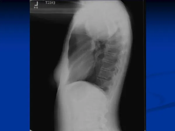
Imaging modalities • Chest X ray • Presence or distortion of mediastinal reflections is the key to interpretation
Imaging modalities • Computed tomography (CT) • Multiplanar reformation images display the detailed anatomical relationship of the tumour with the adjacent structures
Imaging modalities • Magnetic resonance imaging (MRI) • Assessment of preoperative relationships with the pericardium, heart cavities, spinal cord and vascular involvement • Chemical-shift MRI - Useful in distinguishing normal thymus and thymic hyperplasia from thymic neoplasms and lymphoma
Our approach • Women And Men >40 Years • Women 10 To 39 Years Of Age • Men 10 To 39 Years Of Age • Children 0 to 9 years Of Age
Women And Men > 40 Years
Journal of Thoracic Oncology 2014 9, S102-S109DOI: (10.1097/JTO.0000000000000294) Copyright © 2014 International Association for the Study of Lung Cancer Terms and Conditions
Most substernal thyroid goiters manifest as- “Hyperdense mediastinal mass in continuity with the thyroid gland and extending posterior to the great vessels on CT”
“Homogeneous or slightly heterogeneous soft tissue mass with smooth or lobular margins is the usual finding” “30% to 50% of patients with thymoma will exhibit paraneoplastic syndromes (Myasthenia gravis, hypogammaglobulinemia and pure red cell aplasia)”
Women 10 To 39 Years Of Age
Journal of Thoracic Oncology 2014 9, S102-S109DOI: (10.1097/JTO.0000000000000294) Copyright © 2014 International Association for the Study of Lung Cancer Terms and Conditions
““B” symptoms such as fever, night sweats, and weight loss” “Enlarged lymph nodes in the cervical, supraclavicular, and axillary regions may be palpable on physical examination”
“Fat or fat–fluid levels is highly suggestive of the diagnosis ( ~50% ) Calcification ~ 25% Structures such as bone or teeth are <10%” “Especially in younger patients (<25 years)”
“Homogeneous or slightly heterogeneous soft tissue mass with smooth or lobular margins is the usual finding” “20-29y + 30-39y ++”
“10 to 19 y, especially in the setting of rapid symptom onset CT ~ A large heterogeneous mass + pleural effusion” “Cytologic analysis is typically sufficient to make the diagnosis”
Men 10 To 39 Years Of Age
Journal of Thoracic Oncology 2014 9, S102-S109DOI: (10.1097/JTO.0000000000000294) Copyright © 2014 International Association for the Study of Lung Cancer Terms and Conditions
“20 - 30y CT ~ A large heterogeneous mass + Lung Metastasis AFP ++ HCG +”
“10 - 20y CT ~ A large heterogeneous mass + Pleural Effusion + B-symptoms LDH +++”
“ Matted, enlarged LNs + Classical B-symptoms WBC + Alk Phosphatase + ”
“Homogenous Mass+Pulm Mets WBC + Alk Phosphatase + ”
Children 0 to 9 years Of Age
Journal of Thoracic Oncology 2014 9, S102-S109DOI: (10.1097/JTO.0000000000000294) Copyright © 2014 International Association for the Study of Lung Cancer Terms and Conditions
“THYMOMA ”
Thymomas • Rare malignant tumors • Account for about 1/2 of anterior mediastinal tumors • 1/3rd of these are associated with myasthenia gravis • Appear as round or oval masses in early stages but irregular shapes with calcifications occurring in later stages • Can invade surrounding structures including mediastinal fat, pleura, major blood vessels and nerves • Fine needle aspiration, core needle biopsy or open biopsy is used to obtain tissue diagnosis