
NIOSOMES
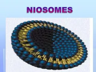
NIOSOMES. They re presents a structure similar to liposome and hence they can represent alternative vesicular systems with respect to liposomes. a) Niosomes are used as an alternative to liposomes, which exhibit certain disadvantages such as : They are expensive

NIOSOMES
E N D
Presentation Transcript
They represents a structure similar to liposome and hence they can represent alternative vesicular systems with respect to liposomes. • a) Niosomes are used as an alternative to liposomes,which exhibit certain disadvantages such as : • They are expensive • Their ingredients like phospholipids are chemically unstable because of their predisposition to oxidative degradation • They require special storage and handling • Purity of natural phospholipids is variable.
b) Differences in characteristics exist between liposomes and niosomes, especially since niosomes are prepared from uncharged single-chain surfactant and cholesterol whereas liposomes are prepared from neutral or charged double-chain phospholipids
Method of preparation In niosomes, the vesicles forming lipid is a non-ionic surfactant such as Span 60 which is stabilized by addition of cholesterol and small amount of anionic surfactant such as dicetyl phosphate. A. Ether injection method • a Mixture of Span 60 , cholesterol and dicetyl phosphate slowly dissolved in diethyl ether then injected slowly through a needle in to warmaqueous phase maintained at 60 °Cthatconsisting of drug. • Vaporization of ether leads to formation of unillamellar niosomes
B. Thin film hydration technique • Mixture of Span 60 , cholesterol and dicetyl phosphateare dissolved in a volatile organic solvent (chloroform) in a round bottom flask. • The organic solvent is removed at room temperature (20°C) using rotary evaporator leaving a thin layer of solid mixture deposited on the wall of the flask. • The dried surfactant film can be rehydratedwith aqueous phase at 60°C with gentle agitation. • This process forms typical multillamellar niosomes.
C. Sonication • Drug solution in phosphat buffer is added to the Mixture of Span 60 , cholesterol and dicetyl phosphate • The mixture is probe sonicated at 60°C for 3 minutes which lead to formation of unillaminarniosomes
Separation of Unentrapped Drug The removal of unentrapped solute from the vesicles can be accomplished by : Dialysis Gel Filtration Centrifugation.
the properties of niosomes depends on the composition of the bilayer: • As the concentration of cholesterol increases, entrapment efficiency decreases. • The entrapment efficiency increases with increase in the concentration and lipophilicity of surfactant. • As HLB value of surfactant decreased give highestpercent entrapment, that Span 60 (HLB = 4.7) gave highest percent entrapment than Span 85 (HLB = 9.8)
Pharmaceutical Applications • Targeting • Sustained Release • Localized Drug Action • since their size and low penetrability through epithelium keeps the drug localized at the site of administration. results in enhancement of efficacy and reduces its systemic toxic effects
PRONIOSOMES • niosomes can forms from proniosomesby coating a water-soluble carrier such as sorbitol or maltodextrin with surfactant. • Where the mixture of maltodextrin and surfactant is dried to form a free flowing powder, in which each water-soluble particle is covered with a thin film of dry surfactant. This preparation is termed “Proniosomes”. • The niosomes are produced by the rehydration of Proniosomes by addition of warm water at T > Tc and brief agitation.
TRANSFERSOME • Transfersomes are complex vesicles that have • extremely flexible & self-regulating membranes, which makes the vesicle very deformable. • Transfersome vesicle can cross microporous barriers efficiently, even if the porous are much smaller than the vesicles size. • Transfersome consists of natural phospholipids suspended in a water-buffered solution containing drug & biocompatible surfactants (sodium cholate). • Similar to a liposome, a Transfersome has a lipid bilayer that surrounds an aqueous core.
The difference between Liposomes & Transfersomes Liposomes are made of phospholipids and to improve the stability of such vesicles, cholesterol is included in the bilayer as membrane (stiffening agent) which lead to more rigid, less flexible and less permeable lipid bilayers. The liposome that applied locally have crossed the skin barrier in a low transport rate and distributed between the cells in building blocks (ceramic layer). The liposome too large to enter the blood vessels; locally they are utilized in peripheral tissues below the application site
Mechanism of Transfersome penetration: The skin is a nanoporous barrier that only permit the passage of smaller particles. Thus the passage of a Transfersome across the skin is due to vesicle membrane flexibility, hydrophilicity, and the ability to perforate the skin barrier.
Explanation of High efficiency of Transfersome transport across the skin compared to liposomes (rigid vesicles) • Ultradeformable, lipid vesicle penetrating a narrow pore, owing to the bilayer components.
When a suspension of Transfersome vesicles is placed on the surface of the skin, the water evaporates from the skin surface and the vesicles start to dry out. • Due to the strong hydrophilicity of Transfersome ingredients, the vesicles are attracted to the areas of higher water content in the narrow gaps between adjoining cells in the skin.
The phenomenon, together with the vesicle's • extreme ability to deform, enables Transfersomes • to change their shape, fit the channels, move across the skin barrier and reach regions of high water content in the deeper skin layers. • Thus, Transfersomes bypass the cutaneous capillary and reach the subcutaneous tissue and the vesicle arrive into the systemic blood circulation.