The Cell Cycle and How Cells Divide
250 likes | 270 Vues
Explore the intricate process of cell division, including DNA synthesis, growth phases, and mitosis, which ensures genetic continuity and reproduction in living organisms.

The Cell Cycle and How Cells Divide
E N D
Presentation Transcript
Phases of the Cell Cycle • The cell cycle consists of • Interphase – normal cell activity • The mitotic phase – cell divsion INTERPHASE (DNA synthesis) Growth G 1 Growth G2 Cell Divsion
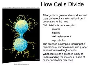
100 µm 200 µm 20 µm (a) Reproduction. An amoeba, a single-celled eukaryote, is dividing into two cells. Each new cell will be an individual organism (LM). (b) Growth and development. This micrograph shows a sand dollar embryo shortly after the fertilized egg divided, forming two cells (LM). (c) Tissue renewal. These dividing bone marrow cells (arrow) will give rise to new blood cells (LM). Functions of Cell Division
Cell Division • An integral part of the cell cycle • Results in genetically identical daughter cells • Cells duplicate their genetic material • Before they divide, ensuring that each daughter cell receives an exact copy of the genetic material, DNA
Figure 12.3 50 µm DNA • Genetic information - genome • Packaged into chromosomes
DNA And Chromosomes • An average eukaryotic cell has about 1,000 times more DNA then an average prokaryotic cell. • The DNA in a eukaryotic cell is organized into several linear chromosomes, whose organization is much more complex than the single, circular DNA molecule in a prokaryotic cell
Chromosomes • All eukaryotic cells store genetic information in chromosomes. • Most eukaryotes have between 10 and 50 chromosomes in their body cells. • Human cells have 46 chromosomes. • 23 nearly-identical pairs
Structure of Chromosomes • Chromosomes are composed of a complex of DNA and protein called chromatin that condenses during cell division • DNA exists as a single, long, double-stranded fiber extending chromosome’s entire length. • Each unduplicated chromosome contains one DNA molecule, which may be several inches long
Maternal set of chromosomes (n = 3) 2n = 6 Paternal set of chromosomes (n = 3) Two sister chromatids of one replicated chromosome Centromere Two nonsister chromatids in a homologous pair Pair of homologous chromosomes (one from each set) Chromosomes • A diploid cell has two sets of each of its chromosomes • A human has 46 chromosomes (2n = 46) • In a cell in which DNA synthesis has occurred all the chromosomes are duplicated and thus each consists of two identical sister chromatids
Homologues • Homologous chromosomes: • Look the same • Control the same traits • May code for different forms of each trait • Independent origin - each one was inherited from a different parent
0.5 µm A eukaryotic cell has multiplechromosomes, one of which is represented here. Before duplication, each chromosomehas a single DNA molecule. Chromosomeduplication(including DNA synthesis) Once duplicated, a chromosomeconsists of two sister chromatidsconnected at the centromere. Eachchromatid contains a copy of the DNA molecule. Centromere Sisterchromatids Separation of sister chromatids Mechanical processes separate the sister chromatids into two chromosomes and distribute them to two daughter cells. Centrometers Sister chromatids Chromosome Duplication • In preparation for cell division, DNA is replicated and the chromosomes condense • Each duplicated chromosome has two sister chromatids, which separate during cell division
Structure of Chromosomes • Diploid - A cell possessing two copies of each chromosome (human body cells). • Homologous chromosomes are made up of sister chromatids joined at the centromere. • Haploid - A cell possessing a single copy of each chromosome (human sex cells).
Phases of the Cell Cycle • Interphase • G1 - primary growth • S - genome replicated • G2 - secondary growth • M -mitosis • C -cytokinesis
Interphase • G1- Cells undergo majority of growth • S - Each chromosome replicates (Synthesizes) to produce sister chromatids • Attached at centromere • Contains attachment site (kinetochore) • G2 - Chromosomes condense - Assemble machinery for division such as centrioles
Mitosis • Some haploid & diploid cells divide by mitosis. • Each new cell receives one copy of every chromosome that was present in the original cell. • Produces 2 new cells that are both genetically identical to the original cell. DNA duplication during interphase Mitosis Diploid Cell
G2 OF INTERPHASE PROMETAPHASE PROPHASE Centrosomes(with centriole pairs) Aster Fragmentsof nuclearenvelope Early mitoticspindle Kinetochore Chromatin(duplicated) Centromere Nonkinetochoremicrotubules Kinetochore microtubule Chromosome, consistingof two sister chromatids Nuclearenvelope Plasmamembrane Nucleolus Mitotic Division of an Animal Cell
METAPHASE ANAPHASE TELOPHASE AND CYTOKINESIS Metaphaseplate Cleavagefurrow Nucleolusforming Nuclear envelopeforming Daughter chromosomes Centrosome at one spindle pole Spindle Mitotic Division of an Animal Cell
G2 OF INTERPHASE Centrosomes(with centriole pairs) Chromatin(duplicated) Nuclearenvelope Plasmamembrane Nucleolus G2 of Interphase • A nuclear envelope bounds the nucleus. • The nucleus contains one or more nucleoli (singular, nucleolus). • Two centrosomes have formed by replication of a single centrosome. • In animal cells, each centrosome features two centrioles. • Chromosomes, duplicated during S phase, cannot be seen individually because they have not yet condensed. The light micrographs show dividing lung cells from a newt, which has 22 chromosomes in its somatic cells (chromosomes appear blue, microtubules green, intermediate filaments red). For simplicity, the drawings show only four chromosomes.
PROPHASE Aster Early mitoticspindle Centromere Chromosome, consistingof two sister chromatids Prophase • The chromatin fibers become more tightly coiled, condensing into discrete chromosomes observable with a light microscope. • The nucleoli disappear. • Each duplicated chromosome appears as two identical sister chromatids joined together. • The mitotic spindle begins to form. It is composed of the centrosomes and the microtubules that extend from them. The radial arrays of shorter microtubules that extend from the centrosomes are called asters (“stars”). • The centrosomes move away from each other, apparently propelled by the lengthening microtubules between them.
METAPHASE Metaphaseplate Centrosome at one spindle pole Spindle Metaphase • Metaphase is the longest stage of mitosis, lasting about 20 minutes. • The centrosomes are now at opposite ends of the cell. • The chromosomes convene on the metaphase plate, an imaginary plane that is equidistant between the spindle’s two poles. The chromosomes’ centromeres lie on the metaphase plate. • For each chromosome, the kinetochores of the sister chromatids are attached to kinetochore microtubules coming from opposite poles. • The entire apparatus of microtubules is called the spindle because of its shape.
ANAPHASE Daughter chromosomes Anaphase • Anaphase is the shortest stage of mitosis, lasting only a few minutes. • Anaphase begins when the two sister chromatids of each pair suddenly part. Each chromatid thus becomes a full- fledged chromosome. • The two liberated chromosomes begin moving toward opposite ends of the cell, as their kinetochore microtubules shorten. Because these microtubules are attached at the centromere region, the chromosomes move centromere first (at about 1 µm/min). • The cell elongates as the nonkinetochore microtubules lengthen. • By the end of anaphase, the two ends of the cell have equivalent—and complete—collections of chromosomes.
TELOPHASE AND CYTOKINESIS Cleavagefurrow Nucleolusforming Nuclear envelopeforming Telophase • Two daughter nuclei begin to form in the cell. • Nuclear envelopes arise from the fragments of the parent cell’s nuclear envelope and other portions of the endomembrane system. • The chromosomes become less condensed. • Mitosis, the division of one nucleus into two genetically identical nuclei, is now complete.
Chromatinecondensing Nucleus Chromosome Nucleolus Metaphase. The spindle is complete,and the chromosomes,attached to microtubulesat their kinetochores, are all at the metaphase plate. 1 2 3 5 Prometaphase.We now see discretechromosomes; each consists of two identical sister chromatids. Laterin prometaphase, the nuclear envelop will fragment. Telophase. Daughternuclei are forming. Meanwhile, cytokinesishas started: The cellplate, which will divided the cytoplasm in two, is growing toward the perimeterof the parent cell. Prophase. The chromatinis condensing. The nucleolus is beginning to disappear.Although not yet visible in the micrograph, the mitotic spindle is staring to from. 4 Anaphase. Thechromatids of each chromosome have separated, and the daughter chromosomesare moving to the ends of cell as their kinetochoremicrotubles shorten. Mitosis in a plant cell
Cytokinesis • Cleavage of cell into two halves • Animal cells • Constriction belt of actin filaments • Plant cells • Cell plate • Fungi and protists • Mitosis occurs within the nucleus