Download
1 / 116
1.18k likes | 1.43k Vues
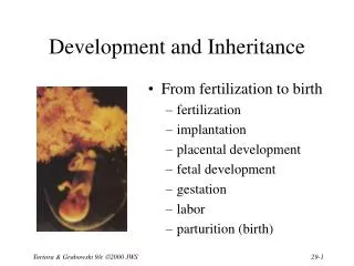
An Introduction to Development and Inheritance. Development Gradual modification of anatomical structures and physiological characteristics from fertilization to maturity Inheritance Transfer of genetic material from generation to generation. 29-1 Development. Differentiation

E N D
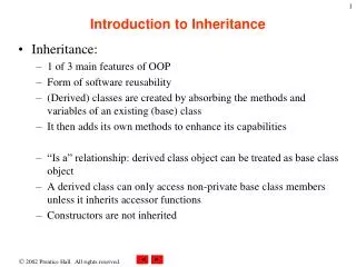
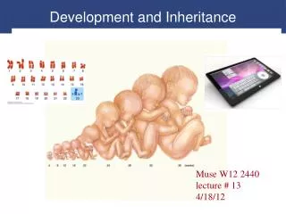
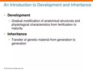
An Introduction to Development and Inheritance • Development • Gradual modification of anatomical structures and physiological characteristics from fertilization to maturity • Inheritance • Transfer of genetic material from generation to generation
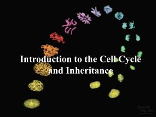
29-1 Development • Differentiation • Creation of different types of cells required in development • Occurs through selective changes in genetic activity • As development proceeds, some genes are turned off, others are turned on • Fertilization • Also called conception • When development begins
29-1 Development • Embryological Development • Occurs during first two months after fertilization • Study of these events is called embryology • Fetal Development • Begins at start of ninth week • Continues until birth
29-1 Development • Prenatal Development • Embryological and fetal development stages • Postnatal Development • Commences at birth • Continues to maturity, the state of full development or completed growth
29-1 Development • Inheritance • Transfer of genetically determined characteristics from generation to generation • Genetics • Study of mechanisms responsible for inheritance
29-2 Fertilization • Fertilization • Fusion of two haploid gametes, each containing 23 chromosomes • Produces zygote containing 46 chromosomes • Spermatozoon • Delivers paternal chromosomes to fertilization site • Travels relatively large distance • Is small, efficient, and highly streamlined
29-2 Fertilization • Gamete • Provides: • Cellular organelles • Inclusions • Nourishment • Genetic programming necessary to support development of embryo for a week
29-2 Fertilization • Fertilization • Occurs in uterine tube within a day after ovulation • Secondary oocyte travels a few centimeters • Spermatozoa must cover distance between vagina and ampulla • Capacitation • Must occur before spermatozoa can fertilize secondary oocyte • Contact with secretions of seminal glands • Exposure to conditions in female reproductive tract
29-2 Fertilization • Acrosomes • Release hyaluronidase and acrosin, enzymes • Penetrate corona radiata, zona pellucida, towardoocyte surface • Oocyte Activation • Contact and fusion of cell membranes of sperm and oocyte • Follows fertilization • Oocyte completes meiosis II, becomes mature ovum
29-2 Fertilization • Polyspermy • Fertilization by more than one sperm • Prevented by cortical reaction • Cortical Reaction • Releases enzymes that: • Inactivate sperm receptors • Harden zona pellucida
29-2 Fertilization • Female Pronucleus • Nuclear material remaining in ovum after oocyte activation • Male Pronucleus • Swollen nucleus of spermatozoon • Migrates to center of cell
29-2 Fertilization • Amphimixis • Fusion of female pronucleus and male pronucleus • Moment of conception • Cell becomes a zygote with 46 chromosomes • Fertilization is complete
29-2 Fertilization • Cleavage • Series of cell divisions • Produces daughter cells • Differentiation • Involves changes in genetic activity of some cells but not others
Figure 29-1a Fertilization A secondary oocyte and numerous sperm at the time of fertilization. Notice the difference in size between the gametes.
Figure 29-1b Fertilization Oocyte at Ovulation Ovulation releases a secondary oocyte and the first polar body; both are surrounded by the corona radiata. The oocyte is suspended in metaphase of meiosis II. Corona radiata First polar body Zona pellucida
Figure 29-1b Fertilization Fertilization and Oocyte Activation Acrosomal enzymes from multiple sperm create gaps in the corona radiata. A single sperm then makes contact with the oocyte membrane, and membrane fusion occurs, triggering oocyte activation and completion of meiosis. Second polar body Fertilizing spermatozoon
Figure 29-1b Fertilization Pronucleus Formation Begins The sperm is absorbed into the cytoplasm, and the female pronucleus develops. Nucleus of fertilizing spermatozoon Female pronucleus
Figure 29-1b Fertilization Spindle Formation and Cleavage Preparation The male pronucleus develops, and spindle fibers appear in preparation for the first cleavage division. Male pronucleus Female pronucleus
Figure 29-1b Fertilization Amphimixis Occurs and Cleavage Begins Metaphase of first cleavage division
Figure 29-1b Fertilization Cleavage Begins The first cleavage division nears completion roughly 30 hours after fertilization. Blastomeres
29-3 Gestation • Induction • Cells release chemical substances that affect differentiation of other embryonic cells • Can control highly complex processes • Gestation • Time spent in prenatal development • Consists of three integrated trimesters, each three months long
29-3 Gestation • First Trimester • Period of embryological and early fetal development • Rudiments of all major organ systems appear • Second Trimester • Development of organs and organ systems • Body shape and proportions change • Third Trimester • Rapid fetal growth and deposition of adipose tissue • Most major organ systems are fully functional
29-4 The First Trimester • First Trimester • Includes four major stages • Cleavage • Implantation • Placentation • Embryogenesis
29-4 The First Trimester • Cleavage • Sequence of cell divisions begins immediately after fertilization • Zygote becomes a pre-embryo, which develops into multicellular blastocyst • Ends when blastocyst contacts uterine wall • Implantation • Begins with attachment of blastocyst to endometrium of uterus • Sets stage for formation of vital embryonic structures
29-4 The First Trimester • Placentation • Occurs as blood vessels form around periphery of blastocyst and placenta develops • Embryogenesis • Formation of viable embryo • Establishes foundations for all major organ systems
29-4 The First Trimester • The First Trimester • Most dangerous period in prenatal life • 40% of conceptions produce embryos that survive past first trimester
29-4 The First Trimester • Cleavage and Blastocyst Formation • Blastomeres • Identical cells produced by cleavage divisions • Morula • Stage after three days of cleavage • Pre-embryo is solid ball of cells resembling mulberry • Reaches uterus on day 4
29-4 The First Trimester • Cleavage and Blastocyst Formation • Blastocyst • Formed by blastomeres • Hollow ball with an inner cavity • Known as blastocoele
29-4 The First Trimester • Cleavage and Blastocyst Formation • Trophoblast • Outer layer of cells separate outside world from blastocoele • Cells responsible for providing nutrients to developing embryo
29-4 The First Trimester • Cleavage and Blastocyst Formation • Inner cell mass • Clustered at end of blastocyst • Exposed to blastocoele • Insulated from contact with outside environment by trophoblast • Will later form embryo
Figure 29-2 Cleavage and Blastocyst Formation Blastomeres Polar bodies 4-cell stage 2-cell stage DAY 2 DAY 1 First cleavage division DAY 0: Fertilization
Figure 29-2 Cleavage and Blastocyst Formation Early morula DAY 3 Advanced morula DAY 4 Hatching Inner cell mass DAY 6 Blastocoele Days 7–10: Trophoblast Implantation in uterine wall (See Figure 29–3) Blastocyst
29-4 The First Trimester • Implantation • Occurs (begins) seven days after fertilization • Blastocyst adheres to uterine lining • Trophoblast cells divide rapidly, creating several layers
29-4 The First Trimester • Implantation • Cellular trophoblast • Cells closest to interior of blastocyst • Syncytial trophoblast • Outer layer • Erodes path through uterine epithelium by secreting hyaluronidase
Figure 29-3 Stages in Implantation DAY 6 FUNCTIONAL ZONE OF ENDOMETRIUM UTERINE CAVITY Uterine glands Blastocyst DAY 7 Trophoblast Blastocoele Inner cell mass
Figure 29-3 Stages in Implantation DAY 8 Cellular trophoblast Syncytial trophoblast DAY 9 Developing villi Amniotic cavity Lacuna
29-4 The First Trimester • Ectopic Pregnancy • Implantation occurs outside uterus • Does not produce viable embryo • Can be life threatening • Lacunae • Trophoblastic channels carrying maternal blood
29-4 The First Trimester • Formation of the Amniotic Cavity • Villi extend away from trophoblast into endometrium • Increase in size and complexity until day 21 • Amniotic Cavity • A fluid-filled chamber • Inner cell mass is organized into an oval sheet two layers thick • Superficial layer faces amniotic cavity • Deeper layer is exposed to fluid contents of blastocoele
29-4 The First Trimester • Gastrulation and Germ Layer Formation • Formation of third layer of cells • Cells in specific areas of surface move toward central line • Known as primitive streak
29-4 The First Trimester • Primitive Streak • Migrating cells leave surface and move between two layers • Creates three distinct embryonic layers, or germ layers • Ectoderm: consists of the superficial cells that did not migrate into interior of inner cell mass • Endoderm: consists of cells that face blastocoele • Mesoderm: consists of poorly organized layer of migrating cells between ectoderm and endoderm
29-4 The First Trimester Ectodermal Contributions Integumentary system: Epidermis, hair follicles and hairs, nails, and glands communicating with the skin (sweat glands, mammary glands, and sebaceous glands) Skeletal system: Pharyngeal cartilages and their derivatives in adults (portion of sphenoid, the auditory ossicles, the styloid processes of the temporal bones, the cornu and superior rim of the hyoid bone)* Nervous system: All neural tissue, including brain and spinal cord
29-4 The First Trimester Ectodermal Contributions Endocrine system: Pituitary gland and adrenal medullae Respiratory system: Mucous epithelium of nasal passageways Digestive system: Mucous epithelium of mouth and anus, salivary glands
29-4 The First Trimester Mesodermal Contributions Integumentary system: Dermis and hypodermis Skeletal system: All components except some pharyngeal derivatives Muscular system: All components
29-4 The First Trimester Mesodermal Contributions Endocrine system: Adrenal cortex, endocrine tissues of heart, kidneys, and gonads Cardiovascular system: All components
29-4 The First Trimester Mesodermal Contributions Lymphatic system: All components Urinary system: The kidneys, including the nephrons and the initial portions of the collecting system Reproductive system: The gonads and the adjacent portions of the duct systems Miscellaneous: The lining of the body cavities (pleural, pericardial, and peritoneal) and the connective tissues that support all organ systems
29-4 The First Trimester Endodermal Contributions Endocrine system: Thymus, thyroid gland, and pancreas Respiratory system: Respiratory epithelium (except nasal passageways) and associated mucous glands Digestive system: Mucous epithelium (except mouth and anus), exocrine glands (except salivary glands), liver, and pancreas
29-4 The First Trimester Endodermal Contributions Urinary system: Urinary bladder and distal portions of the duct system Reproductive system: Distal portions of the duct system, stem cells that produce gametes
29-4 The First Trimester • Embryonic Disc • Oval, three-layered sheet • Produced by gastrulation • Will form body of embryo • Rest of blastocyst will be involved in forming extraembryonic membranes
Figure 29-4 The Inner Cell Mass and Gastrulation Day 10: Yolk Sac Formation Syncytial trophoblast Cellular trophoblast Amniotic cavity Yolk sac Blastocoele Lacunae Superficial layer Deep layer
Figure 29-4 The Inner Cell Mass and Gastrulation Day 12: Gastrulation Yolk sac Amnion Ectoderm Mesoderm Primitive streak Endoderm Blastodisc Embryonic disc