
50 m
50 m. Figure 28.1c Too diverse for one kingdom: a slime mold ( Physarum polychalum ). Figure 28.4 A model of the origin of eukaryotes. Plastid. Dinoflagellates. Alveolates. Apicomplexans. Secondary endosymbiosis. Cyanobacterium. Ciliates. Red algae. Primary endosymbiosis.
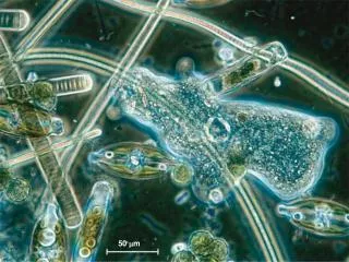

50 m
E N D
Presentation Transcript
Figure 28.1c Too diverse for one kingdom: a slime mold (Physarum polychalum)
Plastid Dinoflagellates Alveolates Apicomplexans Secondary endosymbiosis Cyanobacterium Ciliates Red algae Primary endosymbiosis Stramenopiles Heterotrophic eukaryote Plastid Euglenids Secondary endosymbiosis Green algae Chlorarachniophytes
Chlorophyta Rhodophyta Diplomonadida Animalia Plantae Fungi Euglenozoa Parabasala Radiolaria Cercozoa Amoebozoa (Opisthokonta) Stramenopila Alveolata (Archaeplastida) Fungi Plants Ciliates Diatoms Euglenids Red algae Metazoans Oomycetes Radiolarians Entamoebas Brown algae Parabasalids Diplomonads Chlorophytes Golden algae Kinetoplastids Dinoflagellates Apicomplexans Foraminiferans Gymnamoebas Charophyceans Choanoflagellates Cellular slime molds Chlorarachniophytes Plasmodial slime molds Ancestral eukaryote Unikonta Excavata Chromalveolata Rhizaria
Flagella 0.2 µm Crystalline rod Ring of microtubules
Figure 28.11x Trypanosoma, the kinetoplastid that causes sleeping sickness 9 m
Figure 28.12 A dinoflagellate Flagella 3 µm
CONJUGATION AND REPRODUCTION Two cells of compatible mating strains align side by side and partially fuse. Meiosis of micronuclei produces four haploidmicronuclei in each cell. Three micronuclei in each cell disintegrate. The remaining micro-nucleus in each cell divides by mitosis. MEIOSIS The cells swap one micronucleus. Macronucleus Haploidmicronucleus Compatiblemates Diploidmicronucleus Diploidmicronucleus 8 3 9 1 2 4 5 6 7 MICRONUCLEARFUSION The cellsseparate. The original macro-nucleus disintegrates. Four micronuclei become macronuclei, while the other four remain micronuclei. Two rounds of cytokinesis partition one macronucleus and one micronucleus into each of four daughter cells. Three rounds of mitosis without cytokinesis produce eight micronuclei. Key Micronuclei fuse,forming a diploid micronucleus. Conjugtion Reproduction
Hairy flagellum Smooth flagellum 5 µm
Figure 28.17 Diatoms: Diatom diversity (left), Pinnularia (left)
Blade Stipe Holdfast
Figure 28.21 The life cycle of Laminaria: an example of alternation of generations
radiolarian Axopodia 200 µm
Figure 28.22 Red algae: Dulse (top), Bonnemaisonia hamifera (bottom)
Figure 28.23 Colonial and multicellular chlorophytes: Volvox (left), Caulerpa (right)