Download
1 / 4
40 likes | 51 Vues
This renders the beam parallel into the posterolateral part of the joint, which happens to be accessible for direct puncture. A 22-gauge spinal needle (3 or 5 inches) is put on the pores and skin so the tip is predicted more than the inferior part of the joint. Using the needle concept to be a marker, 510 m, L of 1% lignocaine is injected into the skin and subcutaneous tissue.

E N D
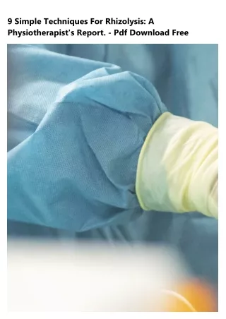
9 Simple Techniques For Rhizolysis: A Physiotherapist's Report. - Pdf Download Free
Rizolisis Cervical Aspect joints are sets of little joints found on either side of your spinal column. They are one of the many structures that keep your spinal column steady and flexible permitting motions like bending and twisting. Sometimes these joints can end up being irritated and this leads to discomfort. Element joints are provided by the median branch nerves and these nerves transmit the pain signals. All 3 treatments require to be performed using X-ray guidance so that we can see the right position of the needle. The treatment is carried out by a Pain Professional in the operating room. Medial Branch Blocks A little amount of
regional anaesthetic is injected near the nerve providing the facet joint. Mielopatia It is a diagnostic block as it will inform us just how much of your pain in the back comes from the facet joints versus other structures. Rhizolysis If deemed ideal following the medial branch blocks, you will have therapeutic median branch rhizolysis at a later date. A radiofrequency needle is put near the medial branch nerves and radiofrequency is utilized to warm the nerves to disrupt their capability to transmit pain signals. Pain decrease can last 12-18 months, for that reason it is our first-line option to treat pain emerging from facet joints. Facet Joint Cortisone Injection A combination of local anaesthetic and steroid is injected around the facet joint to supply discomfort relief. This is the second-line option for dealing with discomfort developing from aspect joints as it is not as long-lasting as rhizolysis although much simpler, simpler and faster to carry out. The Definitive Guide to Radiofrequency Facet Denervation - Bcbsnd The potential adverse effects are: Bleeding and infection Temporary boost in discomfort. This is especially so after rhizolysis and element joint injection. Take regular basic analgesics such as Paracetamol for the first couple of days with application of heat or ice bags. Discomfort at the site of injection. Procedure: The whole treatment will be described to you and the medical professional will ask you to sign a consent kind. In the operating theatre, you will be asked to lie face down on the operating table and the location will be cleaned up with antiseptic. The X-ray machine is utilized to ensure the needle is in the best position near the aspect joint or median branch nerves. Rhizolysis is a term utilized to explain a procedure in which a special probe is put down near to a nerve that comes from the element joints of the back spinal column. By using radio frequency stimulation, the nerve is warmed which stops it sending signals back to the spine. The position of this nerve, which is called the median branch of the posterior primary ramus, is sited at a particular physiological area in the back spine, which can be found under image accumulation (x-ray). The video is not found, possibly removed by the user.
The video is not found, possibly removed by the user. The probe is positioned through the skin, which has been anaesthetised with regional anaesthetic, and when the probe is in the correct position, more local anaesthetic is utilized so that the actual process of lesioning the nerve is less painful. Patients are asked to keep a pain diary describing their symptoms in the first day and the very first, 2nd and third weeks following the treatment, on a scale of 1-10, where 10/10 is the worst discomfort imaginable and 1/10 is a dull ache, taping ratings for Additional reading the back as well as for any leg symptoms that might have been explained prior to the treatment.