
Hookworms
E N D
Presentation Transcript
Hookworms The hookworms cause hookworm disease, which is one of the five major parasitic disease in China(malaria, shistosomiasis, filariasis, kala- azar and hookworm disease). At least two species of hookworms infect man, Necator americanus(美洲板口线虫)and Ancylostoma duodenale(十二指肠钩口线虫). They live in small intestine.
I. Morphology 1. Adults: They look like an odd piece thread and are about 1cm. They are white or light pinkish when living. ♀is slightly larger than♂.The male’s posterior end is expanded to form a copulatory bursa. 2. Eggs: 60×40 µm in size, oval in shape, shell is thin and colorless. Content is 2-8cells.
Differences between two hookworms Adults of A. duodenale Adults of N. americanus
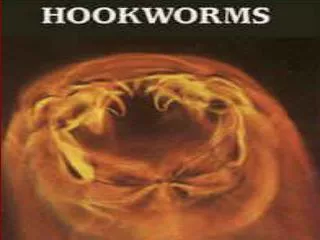
Scanning electron micrograph of the mouth capsule of Ancylostoma duodenale, Note the presence of four "teeth," two on each side.
Scanning electron micrograph of the mouth capsule of Necator americanus, another species of human hookworm. Note the presence of two cutting "teeth“.
Ancylostoma duodenale - copulatory bursa and spines of male(a side view)
Left picture: Copulatory bursa and spines of N. americanus(a side view); • Right picture: copulatory bursa of A. duodenale(a top view)
The bursa of the male canine hookworm (A. caninum) can be seen wrapped around the female hookworm during the act of copulation
Morphologically it is not possible to differentiate between A. duodenale and N. americanus. Interference contrast. ×400. Enlarged by 5.4.
3.The Morphological Differences between Two species of Hookworms _____________________________________________________ A. duodenale N. americanus ______________________________________________________ Size larger smaller ______________________________________________________ Shape single curve, looks like C double curves, looks like S ______________________________________________________ Mouth 2 pairs of ventral teeth 1peir of ventral cutting plates ____________________________________________________________ Copulatory circle in shape oval in shape Bursa (a top view) (a top view) ____________________________________________________________ Copulatory 1pair with separate 1pair of which unite to form spicule endings a terminal hooklet _______________________________________________________ caudal spine present no _______________________________________________________ vulva position post-equatorial pre-equatorial _______________________________________________________
Hookworm egg Decorticated ascaris egg
Differences between Decorticated Ascaris and Hookworm eggs _______________________________________ Decorticated ascaris egg hookworm egg ____________________________________________________________________ Shell thick thin ____________________________________________________________________ Egg cell unsegmented 4-8cells ____________________________________________________________________ Space between new moon shaped space empty space surrounding Shell and cell between cell and ends of shell the segmented cells ____________________________________________________________________
II. Life Cycle 1.Final host: man 2.Inf. Stage: Larva 3 or filariform larva 3.Inf. Route: by skin 4.Food: blood and tissue fluid 5.Site of inhabitation: small intestine 6.Life span: Ad 15years, Na 3-7years 7. Blood-lung migration: skin, cavum, right heart, lungs
8. The differences between the life cycle of Ascaris and Hookworm. ____________________________________________________________ A. lumbricoides Hookworm ____________________________________________________________ Infective stage embryonated egg filariform larvae _____________________________________________________________________ route of infection by mouth by skin _____________________________________________________________________ mode of infection passively actively _____________________________________________________________________ blood-lung pass through the liver don’t pass through the liver migration in lungs of host, larvae the larvae don’t molt and molt twice and stay stay in the lungs for10 days _____________________________________________________________________ food of the adults intestinal content blood _____________________________________________________________________ life span 1 year several years _____________________________________________________________________
Penetrate skin Filariform larvae cavum right heart vesseles, lymphatics lungs(alveolus, bronchiole, bronchus) trachea, pharynx molt3,4 deposit duodenum adults eggs Passed 25-30℃,moisture outside of the body rhabditiform larvea1 In feces O2, 24hours molt1 molt2 rhabditiform larvae2 (L2) 48hrs 6days survive 15weeks in warm soil filariaform(L3) wait for new hosts
III. Pathogenesis and Clinical Manifestations 1. Larval migration (1) Dermatitis, known as "ground itch" or "stool poison".The larvae penetrating the skin cause allergic reaction, petechiae 0r papule with itching and burning sensation. Scratching leads to secondary infection. (2) pneumonitis (allergic reaction), Loeffier's syndrome: cough, asthma, low fever, biood-tinged sputum or hemoptysis, chest-pain, inflammation shadows in lungs under X-ray. These manifestations go on about 2 weeks.
2. Adults in small intestine (1) Epigastric pain as that of a duodenal ulcer. (2) A large worm burden results in microcytic hypochromatic anemia (*character manifestation). The symptoms are lassitude, edema, palpitation of the heart. In severe case, death may result from cardiac failure or physical exhaustion. (3) Allotriophagy (or pica 异嗜症) is due to the lack of trace element iron . (4) Amenorrhea(闭经), sterility(不孕), abortion(流产) may take place in women. (5) Gastrointestinal bleeding (6) Infantile hookworm disease
IV. diagnosis Criterion: 1. hemoglobin is lower than 120g/L in man, 110g/L in woman. 2. find hookworm egg Method: 1. saturated brine flotation technique 2. direct fecal smear 3. culture of larvae V. Treatment 1. Albendazole 2. Mebedazole VI. Epidemiology worldwide distribution. 22-26℃ is the optimal temperature for Ancylostoma duodenale development, Ancylostoma duodenale mainly prevalent in north of China. 31-35℃ is suitable for Necator americanus, it is mainly prevalent in south of China VII. Prevention Unified measures: 1. sanitary disposal of night soil, 2. individual protection, 3. health education, 4. cultivate hygienic habits, 5. treat the patients and carriers.
Enterobius vermicularis The pinworms are one of the most common intestinal nematodes. The adult worms inhabit the cecum and colon. Right after mating, the male dies. Therefore, the male worms are rarely seen. The female worms migrate out the anus depositing eggs on the perianal skin. Humans get this infection by mouth and by autoinfection.
I. Morphology 1. Adults: The adults look like a pin and are white in color. The female worm measures about 8 to 13 mm in size and is fusiform in shape. The male adult is only 2-5mm. The tail of a male is curved. They die right after mating, thus males are rarely seen. The anterior end tapers and is flanked on each side by cuticular extensions called “ cephalic alae”. The esophagus is slender, terminating in a prominent posterior bulb , which is called esophageal bulb. The cephalic alae and esophageal bulb are important in identification of the species. . 2. Egg: 50 to 60m by 25 µm, persimmon seed-like, colorless and transparent, thick and asymmetric shell, content is a larva.
Anterior part of E. vermicularis. Note cephalic alae and esophageal bulb .
The cephalic alae are clearly seen at the anterior end. The cuticle and the alae are transversely striated. The oesophageal bulb are also visible.
Egg: 50 to 60m by 25 µm, persimmon seed-like, colorless and transparent, thick and asymmetric shell, content is a larva.
Anal smear showing large numbers of Enterobius eggs under the lower power. In the background are also two Ascaris eggs.
II. Life cycle 1. site of inhabitation: cecum and colon 2. infective stage: embryonated egg 3. infective route: by mouth . 4. without intermediate host and reservoir host 5. life span of female adults: 1-2 months migrate out anus 6hours Adults eggs on perianal skin embryonated eggs lay swallowed by hostlarvae hatch out molt intestinal lumen adults in cecum 2-4 wks migrate down
III. Symptomatology About one-third of pinworm-infected persons are asymptomatic, The adult worms may cause slight irritation of the intestinal mucosa. Major symptom is anal pruritus, which associates with the nocturnal migration of the gravid females from the anus and deposition of eggs in the perianal folds of the skin. Restlessness, nervousness, and irritability, probably resulting from poor sleep associated with anal pruritus,. In young girls, migration of the worms may produce vaginitis and salpingitis or granuloma of the peritoneal cavity.
IV. Diagnosis Diagnosis depends on recovery of the characteristic eggs. The eggs and the female adults can be removed from the folds of the skin in the perianal regions by the use of the cellophane tape method. The examination should be made in the morning, before the patient has washed or defecated
V.Treatment and prevention Since the life span of the pinworm is less than two months, the major problem is reinfection. Albendazole is the drug of choice. Repeated retreatment may be necessary for a radical cure. Prevention: 1. treat the patients and carriers 2. individual health 3. public health 4. health education and hygienic habits • VI. Epidemiology Geographical distribution—cosmopolitan in temperate zones with about 30 to 50% of the population infected. It is more common in white than colored people and more prevalent in children than adults. Enterobiasis is most common where people live under crowded conditions such as orphanages, kindergartens, and large families.