Download
1 / 1
20 likes | 88 Vues
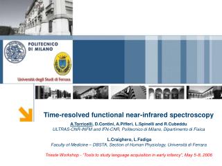
Barrett MD 1 , Cairns SA 1,2 , Whitaker IS 1 , L Hiew 1 , Boyce DE 1 , Cooper MC 1 , Leaper DJ 2 1. Department of Plastic, Reconstructive and Burns Surgery, Welsh Centre for Burns and Plastic Surgery, Swansea, Wales, UK 2. Department of Wound Healing, Cardiff University, Cardiff, UK.

E N D
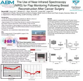
BarrettMD1, Cairns SA1,2, Whitaker IS1, L Hiew1, Boyce DE1, Cooper MC1, Leaper DJ2 1. Department of Plastic, Reconstructive and Burns Surgery, Welsh Centre for Burns and Plastic Surgery, Swansea, Wales, UK 2. Department of Wound Healing, Cardiff University, Cardiff, UK The Use of Near-Infrared Spectroscopy (NIRS) for Flap Monitoring Following Breast Reconstruction After Cancer Surgery • Introduction • In the UK, following mastectomy patients are offered a choice of reconstructive options including myocutaneous flaps. • Approximately 3-5% of these flaps fail due to vascular embarrassment, and require surgical intervention, leech therapy or flap removal. • Often clinical signs of failure appear too late and using a repeatable, non-invasive, real-time system that differentiates between venous and arterial occlusion is invaluable. • Prospective, non-randomized, observational study. Results Nineteen patients completed the study. Fourteen had a normal post-operative course with no significant changes in StO2 or THI. One patient showed a downwards trend in their StO2 with normal clinical observations. In five patients the monitor gave an ‘early warning’ to impending flap compromise, manifested post-operatively with an upward trend in THI, preceding clinical signs of flap failure by up to two hours. Two of these patients required haematoma evacuation with flap survival. three patients required flap removal after several attempts to revise the venous anastamosis. St02 Direct measurement of haemoglobin oxygen in the microcirculation of tissue • THI (Tissue Haemoglobin Index) • A relative index index of the amount of haemoglobin in the volume of tissue illuminated by the sensor. • May differentiate between venous and arterial occlusion Method Twenty patients undergoing free and pedicle myocutaneous flap breast reconstruction, either as an immediate or delayed reconstruction were enrolled. Using the Hutchinson Inspectratm StO2 Tissue Oxygenation Monitor, measurements of tissue oxygen saturation (StO2) and Tissue Haemoglobin Index (THI) were recorded pre-operatively, intra-operatively and continuously post-operatively for 72 hours. Clinical observations were also recorded. Data were correlated with clinical outcome measures. Discussion Near infra-red spectroscopy (NIRS) has potential in the field of reconstructive surgery and the Inspectra StO2 monitor has proved to be a useful, non-invasive monitoring tool. Whilst not replacing the need for experienced and attentive nursing care, we believe it can be a valuable and objective means of assessing flap viability. Successful flap Unsuccessful flap