Download
1 / 61
670 likes | 961 Vues
This document highlights the mechanisms and consequences of blunt ocular trauma, detailing how injuries to the anterior and posterior segments can lead to varied visual prognosis. It discusses complications such as hyphema, corneal opacity, traumatic mydriasis, and cataracts resulting from such trauma. The understanding of injury mechanics, including coup and contrecoup damages, helps in diagnosing and managing these conditions effectively. Key treatments for acute injuries and long-term care strategies emphasize the importance of monitoring changes over time to ensure optimal visual health outcomes.

E N D
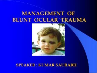
BLUNT OCULAR TRAUMA Dr Soujanya K
general rule anterior segment, or the posterior half is preferentially affected.
Delayed or progressive changes: in some cases • Guarded visual prognosis for all cases • Review for months to years
Coup or Direct damage max. damage at the site of impact
Contre-coup damage max. damage at a point distant from the actual site of impact
Blunt injury Mechanism of injury Dr UmaKulkarni
Blunt injury Mechanism of injury Dr UmaKulkarni
Blunt injury Inside-out injury Mechanism of injury Dr UmaKulkarni
A, direct impact; B, compression wave force; C, reflected compression wave; D, rebound compression wave.
subconjunctivalhaemorrhage: no treatment.
CORNEA Abration > distortion of corneal reflex > flouresceinstain > acute pain & lacrimation
RECURRENT EROSIONS -Spontaneous or injury with babies’ fingernails. -Heals and recurs after days,weeks or months. - occurs on waking up in the morning - Epithelium loosely attached to the Bowmans membrane
Treatment >Debridethe loose epithelium and > pad the eye for 48hrs or bandage soft contact lens.
Blood Staining Of Cornea >colourdep [reddish brown or greenish ]-On duration >clears slowly from periphery
CORNEAL OPACITY • Stromal edema • DM folds Stromal edema, DM Folds DM Tear
Sclera • Partial thickness scleral wounds (lamellar scleral lacerations)
Iris Traumatic miosis
TRAUMATIC MYDRIASIS Large and immobile pupil minute ruptures in the pupillary margin
IRIDODIALYSIS • Black biconvex area at the periphery • D shaped pupil • Distant direct O’scopy- red glow in the periphery • Zonules may also be seen • Monocular Diplopia is a common complaint
ANTEFLEXION OF THE IRIS Pigmented back of the iris faces forwards
Retroflexion of the iris • Whole of the iris is doubled back into the ciliary region and becomes invisible.
ANGLE RECESSION GLAUCOMA • Traumatic secondary open angle glaucoma • The ciliary body is torn- • longitudinal muscle remains attached to the spur at its insertion • circular muscle, pars plicata and the iris root are displaced posteriorly
Clinical features • Unilaterally raised IOP • Abnormally deep AC in the involved meridian • Hyhaema • Gonioscopy- Irregularly broad CB bandassociated with changes of optic neuropathy.
Circular ring of brown pigment –on ant. capsule. • Due to striking of the contracted pupillary margin against the crystalline lens. • It is always smaller than the size of the pupil.
Concussion cataract • Imbibition of aqueous • Direct mechanical
Early Rosette Cataract Feathery lines of opacities along the star-shaped suture lines; usually in the posterior cortex
LATE ROSETTE CATARACT • Posterior cortex, 1 to 2 years after the injury. • Its sutural extensions are shorter and more compact
Discrete subepithelial opacities • Traumatic zonular cataract. • Early maturation of senile cataract • Traumatic absorption of the lens